

Research into peptides for muscle development and recovery has expanded significantly over the past decade. These short chains of amino acids interact with specific cellular receptors to influence various physiological processes, from growth hormone secretion to tissue repair mechanisms. As laboratory research continues to explore their mechanisms of action, understanding the distinct categories of muscle-focused peptides and their documented effects becomes essential for informed scientific investigation.
Understanding Peptides and Their Role in Muscle Physiology
Peptides are biological molecules consisting of two or more amino acids linked by peptide bonds. Unlike proteins, which contain hundreds or thousands of amino acids, peptides are shorter sequences that can trigger specific cellular responses. When researchers examine peptides for bodybuilding and muscle development, they typically focus on two primary categories: growth hormone secretagogues and tissue repair peptides.
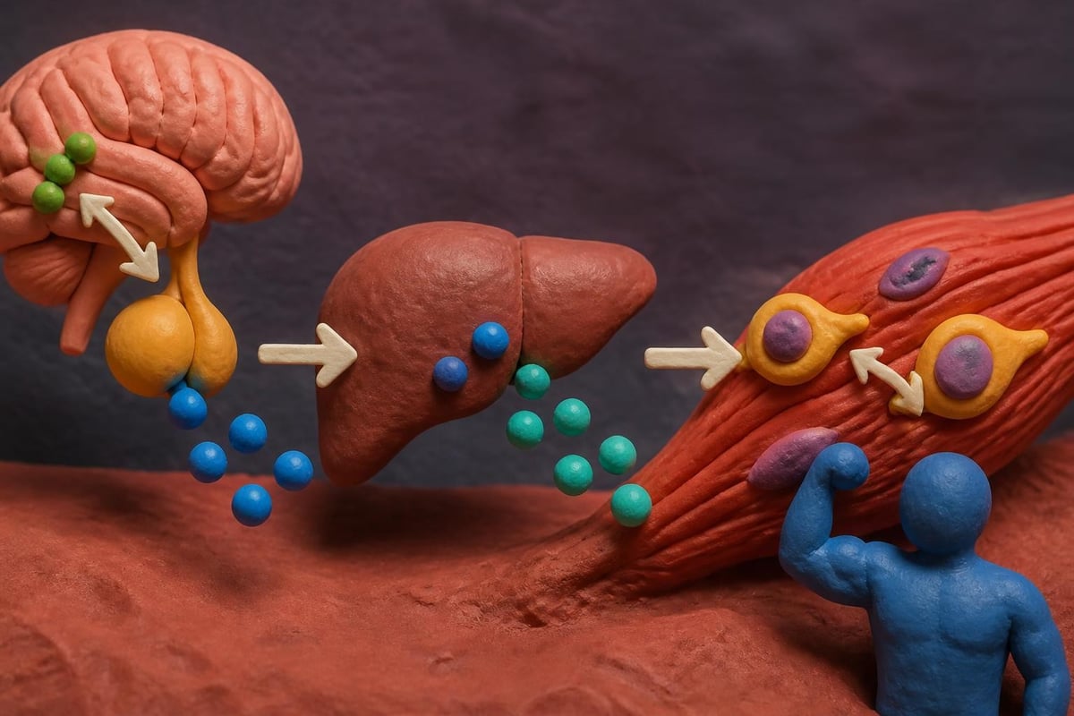
Growth hormone secretagogues work by stimulating the pituitary gland to increase endogenous growth hormone production. This indirect approach differs from synthetic growth hormone administration. By triggering the body's natural production pathways, these peptides maintain more physiological patterns of hormone release.
Tissue repair peptides operate through different mechanisms. They influence cellular migration, angiogenesis, collagen synthesis, and inflammatory modulation. These peptides interact with various receptors throughout the body to accelerate healing processes in muscles, tendons, and connective tissues.
Mechanisms of Action in Muscle Tissue
The biological activity of peptides for muscle depends on their specific amino acid sequences and target receptors. Growth hormone-releasing peptides bind to ghrelin receptors in the pituitary gland and hypothalamus, triggering a cascade that results in growth hormone secretion. This increased growth hormone then promotes insulin-like growth factor 1 (IGF-1) production in the liver.
IGF-1 plays a crucial role in muscle protein synthesis and satellite cell activation. These satellite cells are muscle stem cells that contribute to muscle fiber repair and hypertrophy following mechanical stress or damage.
Research peptides that focus on tissue repair utilize different pathways:
- Fibroblast activation: Stimulating cells responsible for collagen and extracellular matrix production
- Angiogenic signaling: Promoting new blood vessel formation to improve nutrient delivery
- Inflammatory modulation: Balancing pro-inflammatory and anti-inflammatory cytokines
- Growth factor upregulation: Increasing production of vascular endothelial growth factor (VEGF) and other repair signals
Growth Hormone Secretagogues in Muscle Research
Growth hormone-releasing peptides (GHRPs) and growth hormone-releasing hormone (GHRH) analogs represent the most extensively studied peptides for muscle in the secretagogue category. These compounds influence growth hormone production through complementary mechanisms, with some researchers investigating their combined effects.
GHRP Mechanisms and Characteristics
GHRPs function as ghrelin receptor agonists. When they bind to these receptors, they trigger intracellular signaling cascades that result in growth hormone release from somatotroph cells in the anterior pituitary. Unlike GHRH analogs, GHRPs can function even when GHRH receptors are downregulated or less responsive.
Different GHRPs exhibit varying selectivity and potency:
| Peptide Type | Receptor Selectivity | GH Release Pattern | Additional Effects |
|---|---|---|---|
| First Generation | Moderate | Pulsatile with some variation | May stimulate appetite, cortisol |
| Second Generation | High | Strong pulsatile | Minimal appetite stimulation |
| Third Generation | Very High | Sustained pulsatile | Highly selective for GH |
The pulsatile nature of growth hormone release matters significantly. Natural growth hormone secretion follows a circadian rhythm with multiple pulses throughout the day, particularly during deep sleep. Peptides that maintain this pulsatile pattern may better preserve physiological feedback loops.
GHRH Analogs and Their Applications
GHRH analogs work by binding to GHRH receptors on pituitary somatotrophs. Modified versions of natural GHRH have been developed to resist enzymatic degradation, extending their half-life and duration of action. These modifications typically involve substituting specific amino acids that are vulnerable to dipeptidyl peptidase-4 (DPP-4) degradation.
When examining scientific evidence on peptides and muscle building, researchers note that GHRH analogs produce a more physiological growth hormone response compared to synthetic growth hormone administration. This approach maintains negative feedback regulation through somatostatin, preventing excessive or prolonged elevation of growth hormone levels.
Some laboratory studies combine GHRH analogs with GHRPs to achieve synergistic effects. This combination approach capitalizes on two separate signaling pathways, potentially amplifying growth hormone release while maintaining natural regulatory mechanisms.
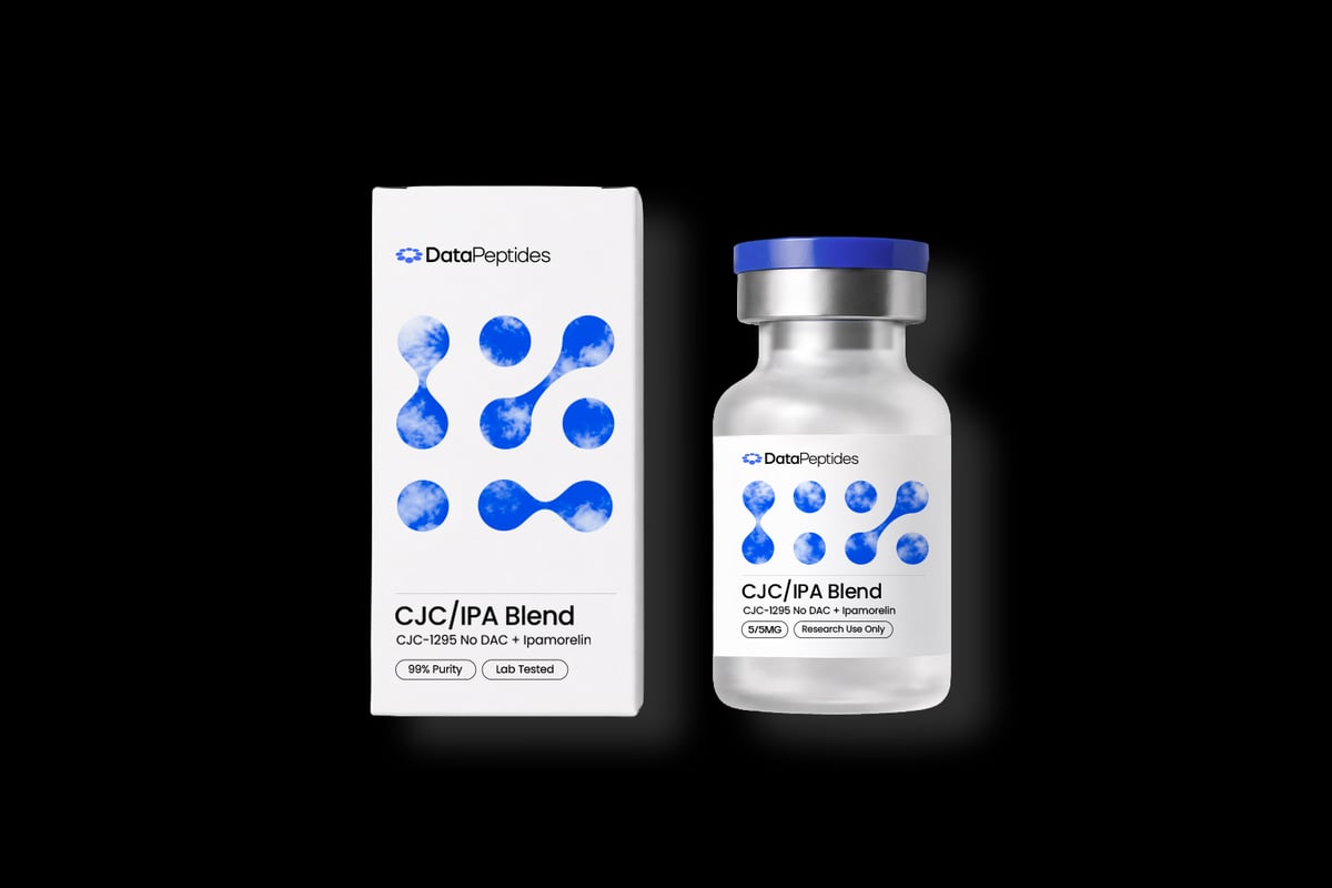
For researchers investigating synergistic approaches, the CJC / IPA Blend offers a combination of a GHRH analog with a selective GHRP, designed to work through complementary pathways for controlled growth hormone elevation in laboratory settings.
Tissue Repair Peptides and Recovery Mechanisms
Beyond growth hormone secretagogues, another category of peptides for muscle focuses specifically on tissue repair, inflammation modulation, and structural healing. These peptides interact with different receptors and cellular pathways compared to secretagogues.
BPC-157 Research Findings
Body Protection Compound 157 is a pentadecapeptide derived from a protective protein found in gastric juice. Laboratory research has explored its effects on various tissue types, including muscles, tendons, ligaments, and the gastrointestinal tract. BPC-157 appears to influence multiple pathways simultaneously.
Studies suggest BPC-157 may:
- Enhance growth factor expression, particularly VEGF
- Modulate nitric oxide production for improved blood flow
- Stabilize cellular structures during stress or injury
- Interact with FAK-paxillin pathway for cell migration
- Influence collagen formation during tissue remodeling
The peptide's stability represents a significant advantage for research purposes. Unlike many bioactive peptides that degrade rapidly in gastric acid or enzymatic environments, BPC-157 maintains structural integrity across various pH levels and temperatures.
TB-500 and Actin Regulation
TB-500 is a synthetic version of Thymosin Beta-4, a naturally occurring peptide present in nearly all human cells. Its primary mechanism involves actin regulation. Actin is a crucial protein for cellular structure, movement, and division. By binding to actin monomers (G-actin), TB-500 influences cell migration and tissue organization.
Research on peptides for recovery highlights TB-500's role in several processes:
- Cellular migration: Enabling cells to move toward injury sites
- Differentiation: Supporting stem cell development into specialized tissue
- Angiogenesis: Promoting new blood vessel formation in damaged areas
- Inflammation control: Modulating inflammatory cytokine production
The peptide's effects on endothelial cells particularly interest researchers studying muscle recovery. Improved vascularization enhances nutrient delivery and waste removal, creating an optimal environment for tissue repair.

Copper Peptides and Structural Support
GHK-Cu represents a copper-binding tripeptide that naturally occurs in human plasma, saliva, and urine. Its concentration decreases with age, which has prompted research into its potential role in tissue maintenance and repair. The copper ion component proves essential for the peptide's biological activity.
GHK-Cu influences multiple aspects of tissue repair:
- Stimulates collagen and glycosaminoglycan synthesis
- Promotes angiogenesis through VEGF and other growth factors
- Functions as an antioxidant to protect against free radical damage
- Modulates metalloproteinase activity for tissue remodeling
- Supports fibroblast migration and proliferation
For muscle tissue specifically, GHK-Cu's effects on extracellular matrix (ECM) organization matter considerably. The ECM provides structural support, transmits mechanical forces, and creates a scaffold for cellular attachment and growth.
Collagen Peptides and Muscle Recovery
Collagen peptides represent a distinct category in peptides for muscle research. These bioactive peptides result from enzymatic hydrolysis of collagen protein, breaking it into smaller, absorbable fragments. Unlike the signaling peptides discussed previously, collagen peptides primarily serve as building blocks for tissue structure.
Bioavailability and Distribution
Studies on collagen peptide supplementation and muscle damage recovery demonstrate that these peptides are absorbed intact in the small intestine. Specific dipeptides and tripeptides enter the bloodstream and accumulate in connective tissues. Research using labeled amino acids has tracked these peptides to tendons, ligaments, and cartilage.
The distribution pattern suggests collagen peptides concentrate where collagen-rich tissues require structural support. This targeted accumulation may explain their effects on connective tissue integrity surrounding muscle fibers.
Mechanisms Supporting Muscle Function
While collagen peptides don't directly build muscle protein (which consists primarily of contractile proteins like actin and myosin), they support the structural framework that enables muscle function:
| Structural Component | Collagen's Role | Impact on Muscle Function |
|---|---|---|
| Endomysium | Surrounds individual muscle fibers | Force transmission, fiber protection |
| Perimysium | Encases muscle fascicles | Coordinate contraction, maintain structure |
| Epimysium | Covers entire muscle | Connect to tendons, distribute mechanical load |
| Tendons | Attach muscle to bone | Transfer contractile force efficiently |
Research indicates that specific collagen peptides combined with resistance training may influence recovery markers and biomechanical characteristics. The peptides appear to support the synthesis and organization of the extracellular matrix that holds muscle tissue together.
Recovery From Exercise-Induced Damage
Eccentric exercise and high-intensity training create microtrauma in muscle fibers and surrounding connective tissue. This damage triggers inflammatory responses and repair processes. Clinical trials examining collagen peptides and exercise-induced muscle soreness have measured markers of muscle damage, inflammation, and functional recovery.
Some findings suggest collagen peptide administration may:
- Reduce markers of muscle damage like creatine kinase
- Modulate inflammatory cytokines during recovery periods
- Support joint comfort during intense training phases
- Enhance structural protein synthesis in connective tissues
Additional research on muscle damage, inflammation, and bone turnover following exercise provides context for understanding how different peptide types might complement each other in comprehensive recovery protocols.
Research Considerations and Laboratory Applications
When designing studies involving peptides for muscle, researchers must account for numerous variables that influence outcomes. These include dosing protocols, timing relative to training or injury, combination approaches, and measurement methodologies.
Dosing and Administration Variables
Peptide research requires precise dosing based on molecular weight, potency, and desired endpoints. Growth hormone secretagogues typically operate in microgram ranges, while tissue repair peptides may require different concentrations depending on the study design.
Researchers consider several factors when establishing protocols:
- Frequency: Single daily administration versus multiple doses
- Timing: Relationship to feeding, training, or sleep cycles
- Duration: Acute studies versus extended protocols
- Route: Subcutaneous, intramuscular, or other delivery methods
- Cycling: Continuous administration versus intermittent patterns
The half-life of each peptide influences these decisions. Some peptides clear rapidly and may require multiple daily administrations to maintain stable levels, while modified versions with extended half-lives allow less frequent dosing.
Combination Approaches in Research
Many investigators explore synergistic effects by combining peptides with complementary mechanisms. For example, pairing a growth hormone secretagogue with a tissue repair peptide addresses both systemic hormone optimization and local healing processes.
Evidence-based comparisons of peptides for muscle growth often discuss these combination strategies:
- GH secretagogue + repair peptide: Systemic anabolic environment plus targeted tissue healing
- Multiple repair peptides: Addressing different aspects of the healing cascade
- Peptides + nutritional interventions: Supporting endogenous processes with substrate availability
- Peptides + training protocols: Investigating timing relative to mechanical stimulus
Each combination requires careful consideration of potential interactions, overlapping pathways, and cumulative effects on measured outcomes.
Measurement and Analysis Methods
Rigorous research on peptides for muscle employs multiple assessment techniques to capture different aspects of muscle physiology and function:
Biochemical markers include growth hormone levels, IGF-1 concentrations, inflammatory cytokines, creatine kinase, and markers of collagen synthesis like procollagen propeptides.
Imaging techniques such as MRI, ultrasound, and DEXA scans provide data on muscle cross-sectional area, muscle quality, and composition changes over time.
Functional assessments measure strength, power output, endurance capacity, and performance in specific tasks relevant to the research question.
Histological analysis of tissue samples (when applicable) reveals cellular changes, fiber type distribution, satellite cell activity, and structural characteristics at the microscopic level.
Quality and Purity Considerations in Peptide Research
The reliability of peptide research depends fundamentally on the quality of the compounds being studied. Peptide synthesis, purification, and storage all influence the actual substance being investigated.
Manufacturing Standards and Testing
High-quality research peptides undergo rigorous synthesis processes, typically solid-phase peptide synthesis (SPPS) or liquid-phase synthesis. After synthesis, purification removes truncated sequences, deletion peptides, and other impurities.
Third-party testing verifies:
- Purity percentage: Proportion of the desired peptide versus contaminants
- Sequence accuracy: Confirming correct amino acid order
- Molecular weight: Mass spectrometry validation
- Sterility: Absence of microbial contamination
- Endotoxin levels: Particularly important for certain research applications
Researchers at Data Peptides can access laboratory-grade compounds that undergo batch testing to verify these quality parameters, ensuring consistency across studies.
Storage and Stability
Peptide stability varies based on sequence composition, formulation, and storage conditions. Most research peptides require specific temperature ranges and protection from light, oxidation, and repeated freeze-thaw cycles.
Proper handling includes:
- Lyophilized storage at appropriate temperatures (often -20°C or -80°C)
- Reconstitution with sterile, appropriate vehicles
- Aliquoting to avoid multiple freeze-thaw cycles
- Protection from light exposure for photosensitive peptides
- Monitoring for signs of degradation or precipitation

Temperature excursions or improper storage can lead to peptide degradation, aggregation, or loss of biological activity, compromising research validity.
Current Research Directions and Emerging Applications
The field of peptides for muscle continues to evolve as researchers identify new sequences, mechanisms, and applications. Several emerging areas show promise for expanding our understanding of muscle physiology and recovery.
Mitochondrial Peptides
Mitochondrial-derived peptides represent a newer area of investigation. These peptides are encoded by mitochondrial DNA and influence cellular metabolism, stress resistance, and energy production. Since mitochondria play crucial roles in muscle energy systems and fatigue resistance, these peptides interest researchers studying muscle performance and recovery.
One example is MOTS-c, which appears to influence metabolic regulation and may affect how muscles utilize energy substrates. Research explores its potential effects on exercise capacity and metabolic flexibility.
Tissue-Specific Targeting
Developing peptides with enhanced tissue specificity represents an ongoing research goal. Rather than systemic effects, these modified peptides would preferentially accumulate in or act upon muscle tissue, potentially improving efficacy while minimizing off-target effects.
Strategies include:
- Conjugating peptides to targeting molecules
- Designing sequences that bind muscle-specific receptors
- Utilizing delivery systems that concentrate in muscle tissue
- Exploiting differences in receptor expression between tissues
Computational Approaches to Peptide Research
Advanced computational tools are transforming peptide research. Peptide databases and predictive models enable researchers to analyze structure-activity relationships, predict binding affinities, and design novel sequences with desired properties.
Machine learning approaches to peptide property prediction use transformer-based models to analyze vast datasets of peptide sequences and their biological activities. These tools may accelerate the identification of promising compounds for muscle-related research.
Regulatory and Ethical Considerations
Research involving peptides for muscle occurs within established regulatory frameworks that ensure safety, scientific integrity, and ethical conduct. These considerations shape how investigations are designed, conducted, and reported.
Research Use Specifications
Laboratory-grade research peptides are manufactured and distributed specifically for investigational purposes. They are not approved for human consumption, therapeutic use, or veterinary applications. This distinction matters for:
- Institutional review processes: Studies involving human subjects require appropriate ethical review
- Laboratory protocols: Research must follow established safety and handling procedures
- Documentation: Proper record-keeping of compound sourcing, handling, and disposal
- Regulatory compliance: Adherence to guidelines governing research substances
Researchers must clearly distinguish between laboratory investigation and clinical application, ensuring their work contributes to scientific understanding while respecting regulatory boundaries.
Data Integrity and Reproducibility
The peptide research community increasingly emphasizes reproducibility and transparent reporting. This includes detailed descriptions of:
- Peptide source, purity, and verification methods
- Exact dosing protocols and administration routes
- Subject or model characteristics and selection criteria
- Measurement techniques and analysis methods
- Statistical approaches and raw data availability
These practices enable other researchers to replicate findings, validate conclusions, and build upon existing work, advancing the field's overall understanding of peptides for muscle applications.
Understanding peptides for muscle requires examining multiple categories of compounds, diverse mechanisms of action, and rigorous research methodologies. From growth hormone secretagogues to tissue repair peptides and structural collagen fragments, each type offers distinct pathways for investigation into muscle physiology, recovery, and performance. For researchers seeking laboratory-grade compounds with verified purity and US-based manufacturing, Data Peptides provides third-party tested research peptides designed specifically for scientific investigation, supporting the advancement of peptide research with reliable, quality-controlled materials.