

KPV is a three-amino-acid fragment of α-melanocyte-stimulating hormone (Lys-Pro-Val) that suppresses NF-κB signaling and reduces inflammatory cytokine output in intestinal, immune, and skin tissue. The strongest evidence base sits in rodent colitis models, where oral KPV depends entirely on the intestinal transporter PepT1 (SLC15A1) to reach inflamed tissue. Human data exists but stops short of confirmation: a Phase 2 signal in ulcerative colitis never advanced to Phase 3, and the peptide carries no FDA approval for any indication.
For researchers deciding where to spend bench time, three facts matter immediately:
- Preclinical support is deep and mechanistically consistent across DSS, TNBS, and AOM/DSS colitis models
- PepT1 dependence means knockout models eliminate the therapeutic effect entirely, a strong causal signal rarely seen this cleanly in peptide research
- No large-scale human trial has replicated the early clinical signal, so dose-response and long-term safety data in humans remain thin
KPV Peptide Research at a Glance: Oral KPV produced measurable reductions in colitis severity scores in PepT1-dependent mouse models. Transdermal delivery through microneedles combined with iontophoresis increased skin permeation by up to 35-fold over passive diffusion.
TL;DR:
- KPV’s efficacy in animal models relies strictly on PepT1 transport, as knockout studies show the absence of benefit without the transporter.
- Human trial data from decades ago indicated potential benefits in ulcerative colitis, but no modern, large-scale confirmation or regulatory approval exists yet.
- Formulation strategies like nanoparticle encapsulation or microneedle-assisted delivery significantly improve stability and tissue penetration for topical and oral applications.
- Confirming KPV purity requires lot-specific certificates backed by mass spectrometry and HPLC, making sourcing quality essential for reproducible results.
- Future research needs contemporary human pharmacokinetic data, dose-finding studies, and replication of tumorigenesis results to verify clinical potential.
Table of Contents
- What Drives KPV Peptide Research: The Melanocortin and PepT1 Connection
- Evidence Review: What the Preclinical and Human Studies Actually Show
- Delivery Routes and Formulation Strategies for KPV Peptide Research
- Safety, Tolerability, and the Current Regulatory Status
- Designing Reproducible KPV Studies: Assays, Models, and Endpoints
- Sourcing Research-Grade KPV: What a Real Certificate of Analysis Should Show
- Where the KPV Evidence Base Still Falls Short
- Reading the KPV Evidence Without Getting Ahead of It
- Get Research-Grade KPV With Verified Purity and Fast US Shipping
- Key Takeaways
- Sources
- FAQ
What Drives KPV Peptide Research: The Melanocortin and PepT1 Connection
KPV’s biological identity starts with its parent molecule. It is the C-terminal tripeptide of α-MSH, but unlike the full hormone, KPV exerts its anti-inflammatory activity independent of melanocortin receptor binding. That distinction matters for experimental design: you don’t need MC1R expression on your target cells to see an effect, which is why KPV shows activity in enterocytes, macrophages, and keratinocytes that vary widely in melanocortin receptor density.
The core mechanism runs through NF-κB, the transcription factor complex that governs much of the inflammatory cytokine cascade. KPV interferes with the IKK/IκB/p65 signaling axis, limiting nuclear translocation of p65 and, in turn, dampening downstream production of TNF-α, IL-6, and IL-8. Reviews of KPV’s in vitro activity report effects at nanomolar concentrations across multiple cell types, consistent with a potent, receptor-independent anti-inflammatory action rather than a broad cytotoxic or nonspecific effect.
The second half of the mechanism is where KPV gets interesting for drug delivery scientists: PepT1-mediated uptake. PepT1 is a proton-coupled oligopeptide transporter normally responsible for absorbing di- and tripeptides from digested protein in the small intestine. Under inflammatory conditions, PepT1 expression shifts. It becomes upregulated in inflamed colonic epithelium, a tissue that in a healthy gut expresses little of the transporter. That upregulation creates a rare pharmacological opportunity: an oral tripeptide that gets preferentially absorbed exactly where the tissue is inflamed, rather than distributing uniformly across the gut.
The disease-site targeting effect isn’t incidental. When researchers knocked out PepT1 in mouse colitis models, oral KPV’s therapeutic benefit disappeared almost completely. The transporter isn’t just a delivery convenience; it’s the mechanism itself.
This PepT1 dependence has been demonstrated directly. Dalmasso and colleagues showed that oral KPV reduced inflammation and improved histological scores in DSS and TNBS colitis models, and that this benefit required functional PepT1 transport. Remove the transporter, and the peptide might as well be an inert filler.
A few cell populations show consistent KPV responsiveness across the literature:
- Enterocytes — the primary site of PepT1-mediated uptake and the target tissue in colitis models
- Macrophages and other immune cells — show reduced pro-inflammatory cytokine secretion following KPV exposure
- Keratinocytes — relevant for topical and wound-healing applications, where NF-κB inhibition limits local inflammatory signaling independent of gut transport
For labs working across gut health mechanisms more broadly, KPV represents one of the cleaner examples of a transporter-dependent, disease-site-targeted peptide. Most anti-inflammatory peptides distribute broadly and rely on systemic exposure; KPV’s PepT1 dependence gives it a built-in targeting mechanism that oral small molecules rarely achieve without a delivery vehicle.
Evidence Review: What the Preclinical and Human Studies Actually Show
The KPV evidence base breaks into three tiers: strong rodent colitis data, a smaller but mechanistically important cancer model, and a historical human trial that never got confirmed. Each deserves separate scrutiny, because pooling them into a single “KPV works” statement overstates what any one study alone supports.
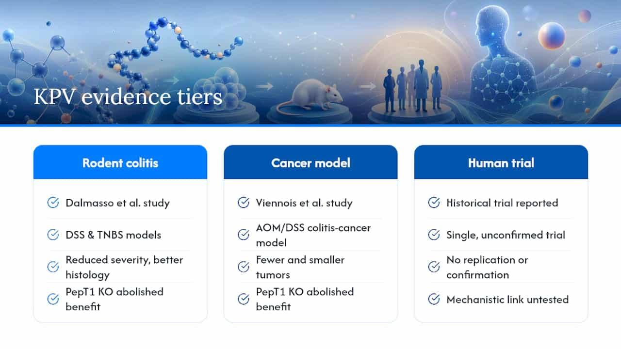
1. Oral KPV in DSS and TNBS colitis models. The foundational study here comes from Dalmasso et al., who administered oral KPV to mice with dextran sodium sulfate (DSS) and trinitrobenzene sulfonic acid (TNBS) induced colitis, two chemically distinct models that together capture different aspects of colonic inflammation. Treated animals showed reduced disease severity and improved histological scoring compared to untreated controls. The critical control experiment used PepT1-knockout mice: without the transporter, oral KPV produced no measurable benefit. That single experiment does more to establish KPV’s mechanism than a dozen dose-response curves would, because it rules out systemic absorption or a PepT1-independent pathway as the source of the effect.
2. Colitis-associated tumorigenesis in the AOM/DSS model. Viennois and colleagues extended the PepT1 story into cancer biology, using the azoxymethane/DSS (AOM/DSS) model, which combines a carcinogen with chronic inflammation to mimic colitis-associated colorectal cancer. KPV-treated mice developed fewer and smaller tumors. Again, PepT1-knockout animals lost the protective effect entirely. This is a meaningful data point for researchers modeling chronic inflammatory disease progression, since it suggests KPV’s benefit isn’t limited to acute inflammation control. It may also blunt the inflammation-to-neoplasia pathway that drives long-term colitis complications, though this remains one dataset rather than a replicated finding across independent labs.
3. Nanoparticle-based colon targeting. The same research group behind the AOM/DSS work also tested hyaluronic acid-functionalized nanoparticles as a delivery vehicle, protecting KPV from degradation in the upper GI tract and concentrating release at inflamed colonic sites. This approach addresses a real formulation problem: free KPV is vulnerable to peptidase degradation before it reaches the colon, and nanoparticle encapsulation improves the odds that an oral dose survives intact to where PepT1 can absorb it.

4. Topical and wound-healing contexts. Outside the gut, KPV’s NF-κB inhibition has translational relevance for skin inflammation and wound repair, where local cytokine suppression can influence healing trajectories. This branch of the literature is thinner than the colitis work and relies more heavily on in vitro keratinocyte data than on validated animal wound models, so treat topical efficacy claims as mechanistically plausible rather than clinically established.
5. Human trial history: Lipotropin-1 and the Phase 2 gap. KPV-based formulations, historically developed under the name Lipotropin-1, produced a Phase 2 signal in ulcerative colitis, with reported improvements in disease activity scores and some evidence of mucosal healing. That program did not advance to Phase 3, and detailed public trial data from that era is limited. This is the single biggest gap in the KPV evidence chain: a human efficacy signal exists, but it was never confirmed at the scale needed for regulatory approval, and the underlying dataset isn’t available for independent reanalysis.
Put together, the evidence hierarchy looks like this: mechanistic plausibility is well established, rodent colitis efficacy is robust and reproduced across two independent research groups, and human confirmation is a single, decades-old, unconfirmed signal. Anyone citing KPV as a “clinically proven” anti-inflammatory therapy is overstating the current record by at least one evidence tier.
Delivery Routes and Formulation Strategies for KPV Peptide Research
Route of administration determines whether KPV’s mechanism can even engage its target tissue, which makes formulation research almost as important as mechanism research for this particular peptide.
Oral dosing exploits the PepT1 pathway directly. In the DSS and TNBS mouse studies, oral gavage delivered KPV successfully to inflamed colonic tissue, but free peptide faces two problems in a real gut environment: enzymatic degradation before absorption and unpredictable release timing. That’s the rationale behind encapsulation strategies.
Nanoparticle and hydrogel colon-targeting systems address the degradation problem directly. Alginate-chitosan and hyaluronic acid-functionalized nanoparticles have been used to shield KPV through the stomach and small intestine, releasing the peptide preferentially at inflamed colonic sites where PepT1 expression is elevated. This is a targeted-release strategy layered on top of a targeted-uptake mechanism, essentially two independent selectivity filters working in sequence.
Transdermal delivery solves a completely different problem. KPV crosses intact skin poorly by passive diffusion alone, which limits its usefulness as a simple topical cream for inflammatory skin conditions. Combining microneedle pretreatment with iontophoresis changes that picture substantially: this approach increased KPV permeation by up to 35-fold compared to passive diffusion, while also improving retention of the peptide within skin layers rather than just surface contact.
- Oral gavage: direct PepT1 engagement, but vulnerable to luminal degradation without encapsulation
- Nanoparticle encapsulation: protects payload and concentrates release at inflamed colonic tissue
- Microneedle-assisted iontophoresis: converts KPV from a poor passive skin penetrant into a viable topical modality
- Free-peptide topical creams: limited evidence of meaningful dermal penetration without an enhancement technique
Pro Tip: If you’re designing a topical KPV protocol, don’t default to a passive-diffusion vehicle and assume it will work because the peptide is small. Microneedle or iontophoretic pretreatment isn’t optional at this molecular scale for meaningful tissue exposure.
Stability remains the recurring headache across every route. KPV is a short peptide, and short peptides are peptidase targets. Cold-chain storage, encapsulation, and careful reconstitution protocols all reduce degradation risk, but researchers should build stability testing into any PK study design rather than assuming shelf stability from a certificate of analysis alone.
Safety, Tolerability, and the Current Regulatory Status
KPV’s tolerability profile in preclinical studies has been favorable, with no major adverse findings reported at doses used in the DSS, TNBS, and AOM/DSS colitis models. The historical Phase 2 human trial data also didn’t flag significant safety signals, though the dataset is limited and decades old, so treat it as reassuring rather than conclusive.
The regulatory picture is unambiguous on one point: KPV is not FDA-approved for any human therapeutic use. It exists in a gray zone common to many bioactive peptides, sold and studied under research-use-only frameworks while its compounding status remains under active review. FDA advisory materials reference ongoing scrutiny of peptide compounding classifications, and the Pharmacy Compounding Advisory Committee’s July 2026 meeting included agenda items specifically addressing peptide compounding rules. That calendar is worth watching for anyone whose research plans depend on future compounded-formulation access, since the classification outcome could change what’s obtainable and under what conditions.
For labs, a few practical points follow from this status:
- KPV should be sourced and used strictly under research-use-only terms, never represented as suitable for human or animal treatment
- Any move toward clinical evaluation requires an IND filing and IRB oversight, and researchers should not assume the historical Lipotropin-1 data will substitute for new safety and pharmacokinetic work
- Sourcing decisions should prioritize suppliers offering documented, lot-specific purity data rather than generic assurances, since regulatory ambiguity makes third-party verification more important, not less
None of this makes KPV a dead end. It means the research pathway runs through standard translational steps: PK characterization, dose-finding, and formal safety studies, the same steps every promising preclinical peptide has to clear before any clinical program can move forward responsibly.
Designing Reproducible KPV Studies: Assays, Models, and Endpoints
Reproducibility problems in peptide research usually trace back to assay selection and endpoint definition rather than the peptide itself. KPV studies benefit from a fairly standardized toolkit at this point.
-
In vitro NF-κB inhibition assays. Reporter gene assays measuring NF-κB transcriptional activity, paired with direct cytokine readouts (TNF-α, IL-6, IL-8 via ELISA or multiplex panels), give the cleanest mechanistic signal. Nuclear p65 translocation can be tracked by immunofluorescence or Western blot fractionation to confirm the IKK/IκB/p65 pathway is the operative mechanism rather than an off-target effect.
-
Colitis model selection. DSS models chemical epithelial damage and innate immune activation; TNBS engages a more T-cell-driven inflammatory response; AOM/DSS adds a carcinogenic component for tumorigenesis endpoints. Choosing the right model, or running more than one, depends on whether the research question is acute inflammation, chronic injury, or cancer progression.
-
Endpoints worth standardizing. Disease activity index, histological injury scoring, colon length (a simple but informative proxy for chronic inflammation severity), and cytokine profiling from colonic tissue homogenate all appear consistently across the published KPV literature. Using the same endpoints as prior studies makes cross-study comparison possible.
-
PK/ADME characterization. This is the biggest methodological gap in the current record. Mass spectrometry-based quantification, particularly LC-MS/MS, remains the most reliable method for tracking KPV concentration in plasma and tissue given the peptide’s short length and rapid clearance profile.
-
Controls and power. PepT1-knockout controls have proven essential for establishing mechanism in prior studies; any oral KPV study should consider including this arm. Underpowered studies with small group sizes are common in peptide pilot work, so pre-registering sample size calculations helps the field avoid another generation of unreplicated positive results.
Sourcing Research-Grade KPV: What a Real Certificate of Analysis Should Show
Reproducibility starts before the experiment, at the point where you decide which vial of KPV to trust. A Certificate of Analysis worth relying on should show lot-specific purity data (ideally 99%+), the analytical method used to establish it, and a date tied to that specific batch, not a generic template reused across products.
Mass spectrometry and HPLC verification are the two workflows that actually confirm identity and purity rather than just asserting it. Mass spec confirms molecular weight matches the expected KPV sequence, while HPLC purity traces show whether degradation products or synthesis byproducts are present above acceptable thresholds. If a supplier can’t produce lot-specific chromatograms or mass spec traces on request, that’s a documentation gap worth taking seriously.
- Confirm the COA lists a specific lot number matching the vial received, not a generic product-level document
- Check that purity is verified by both mass spec (identity) and HPLC (purity), not one method alone
- Store lyophilized peptide at recommended cold-chain conditions and avoid repeated freeze-thaw cycles after reconstitution
- Document chain-of-custody from supplier to bench, especially for studies intended for peer review or regulatory submission
- Avoid consumer-grade sources with no lot-specific testing, generic marketing language, or COAs that predate the batch sold
Pro Tip: Cross-check the COA’s stated molecular weight against the theoretical mass for Lys-Pro-Val (346.4 g/mol as the free base) before you run a single assay. A mismatch here catches sourcing problems before they cost you weeks of misattributed results.
Data Peptides maintains lot-specific Certificates of Analysis for its research peptide catalog, giving labs a documented reference point when qualifying a new supplier or replicating a published protocol. For broader guidance on evaluating research peptide quality before committing to a vendor, a documented QC process matters more than a low price point every time.
Where the KPV Evidence Base Still Falls Short
The honest gap list for KPV is short but consequential. No Phase 3 trial has ever been completed, which means there’s no large-scale, modern human efficacy or safety dataset to point to. The Phase 2 Lipotropin-1 signal from decades ago hasn’t been replicated under current trial standards or with contemporary formulation approaches.
PK and ADME data in humans remains essentially absent. Researchers know a great deal about KPV’s behavior in mouse colon tissue and comparatively little about its half-life, bioavailability, or dose-response relationship in humans, oral or otherwise. Endpoint standardization across the animal literature is decent for colitis models but nonexistent for topical and wound-healing applications, where studies use inconsistent metrics that make cross-study comparison difficult.
Priority next steps for the field:
- Modern human PK studies using mass spec quantification to establish basic bioavailability parameters
- Dose-finding studies that replicate the historical Phase 2 signal under current trial design standards
- Multi-center replication of the AOM/DSS tumorigenesis findings to confirm the anti-neoplastic effect beyond a single research group
- Standardized topical/wound-healing endpoints to make the dermatological evidence base comparable across studies
Reading the KPV Evidence Without Getting Ahead of It
The PepT1 dependence data is, frankly, the most convincing piece of KPV’s entire story, and it’s the part that gets underplayed. Most peptide research settles for correlation. Knockout models that erase the effect entirely are rare, and that’s exactly what happened twice, in two different disease contexts, from two research groups. That’s a real mechanistic anchor, not just a promising trend line.
Where we think the field gets ahead of itself is in treating the Phase 2 Lipotropin-1 signal as if it were a modern, confirmable finding. It’s historical, undetailed by current standards, and unreplicated. Researchers building on KPV should treat human efficacy as an open question requiring new PK work and rigorous dose-finding, not as settled science waiting for a rubber stamp.
The practical path forward for any lab is unglamorous but necessary: characterize pharmacokinetics properly, choose a formulation strategy that matches your delivery goal, and insist on lot-verified material with real chromatography behind it. We built our gut health peptide resources around that same principle. Good research starts with materials you can actually trust and verify, not just a name on a label.
— Data Peptides
Get Research-Grade KPV With Verified Purity and Fast US Shipping
Sourcing ambiguity is the biggest avoidable risk in KPV research, and it’s the one problem Data Peptides was built to solve. Every peptide in the catalog ships with a lot-specific Certificate of Analysis backed by third-party mass spec and HPLC verification, so you’re not guessing whether your vial matches the purity claim on the label. Browse the current peptide catalog to check lot availability and COA data before your next protocol, and if KPV isn’t the right fit for your model, the full research peptide range covers adjacent mechanisms worth comparing.
Key Takeaways
KPV’s anti-inflammatory effect depends on PepT1-mediated transport into inflamed tissue, and human confirmation of its efficacy remains an open research question despite strong preclinical signals.
| Point | Details |
|---|---|
| Mechanism is receptor independent | KPV inhibits NF-κB signaling without requiring melanocortin receptor binding, working across enterocytes, immune cells, and keratinocytes. |
| PepT1 dependence is the key finding | Knockout models eliminated KPV’s benefit in both colitis and tumorigenesis studies, confirming transporter-mediated targeting. |
| Human evidence stops at Phase 2 | The historical Lipotropin-1 trial showed a signal in ulcerative colitis but never advanced to Phase 3 confirmation. |
| Transdermal delivery needs enhancement | Passive skin diffusion is poor; microneedles plus iontophoresis boosted permeation up to 35-fold. |
| Sourcing quality is non-negotiable | Verify lot-specific COAs with mass spec and HPLC data; Data Peptides provides this documentation for its research peptide catalog. |
Sources
- PepT1-Mediated Tripeptide KPV Uptake Reduces Intestinal Inflammation
- Orally targeted delivery of tripeptide KPV via hyaluronic acid–functionalized nanoparticles efficiently alleviates ulcerative colitis
- FDA document (related advisory/regulatory material)
This article is general information, not a substitute for advice from a qualified doctor. Consult a qualified healthcare professional about your own circumstances before acting on anything here.
FAQ
Does KPV Peptide Actually Work?
Preclinical evidence in colitis models is strong and mechanistically well supported through PepT1-dependent uptake, but human confirmation stops at an unreplicated Phase 2 signal, so “works” currently applies more confidently to animal models than to people.
Who Should Avoid KPV Peptide Research Use?
KPV is not FDA-approved for human or animal treatment, so it should be limited to qualified research settings under research-use-only terms rather than any direct consumption context.
How Long Should KPV Be Used in Research Protocols?
Duration depends entirely on the study design. Published colitis models typically dosed KPV over the course of induced-inflammation periods lasting one to two weeks, and no standardized human dosing duration exists given the absence of confirmed Phase 3 data.
What Is the Best KPV Peptide Source for Research?
The best source is one that provides lot-specific Certificates of Analysis backed by mass spec and HPLC verification rather than generic purity claims; Data Peptides publishes this documentation for each batch in its research catalog.