

Peptides can meaningfully accelerate specific stages of wound repair, and the strongest evidence points to cyclic and rationally designed scaffolds rather than simple linear chains. That’s the short answer. The longer answer depends on which wound phase you’re targeting, how well the peptide survives protease-rich wound fluid, and whether anyone has bothered to test it in a diabetic or ischemic model instead of a healthy young mouse.
The mechanistic case is solid. WHP1, a template-designed peptide, drove dose-dependent migration and proliferation in HaCaT keratinocyte scratch assays, protected cells against oxidative stress, and pushed macrophages to secrete more TGF‑β. LL‑37, the best-studied human antimicrobial peptide, does double duty by clearing bacteria while also driving keratinocyte migration through immune and tissue signaling pathways. Cyclic peptides like CyRL‑QN15 bring something linear chains generally can’t: enough protease resistance to survive long enough in the wound bed to matter, with preclinical data showing improved epithelization, collagen deposition, and angiogenesis.
Here’s the honest translational picture:
- Most high-quality peptide data for wound healing sits at the in vitro and small-animal in vivo stage.
- A handful of cyclic or synthetic scaffolds have moved into early development pipelines, but large randomized clinical trials remain rare.
- Delivery format (hydrogel, nanoparticle, electrospun fiber) often determines whether a mechanistically sound peptide survives contact with wound exudate.
- Diabetic and ischemic wound models remain underrepresented relative to standard excisional models, which limits how confidently anyone can predict clinical performance.
The gap in peptide wound healing research isn’t proving a mechanism works in a dish. It’s proving the peptide survives the wound environment long enough for that mechanism to matter in a body with impaired healing.
For researchers starting a project this quarter, the practical move is matching a peptide’s known mechanism to a specific wound phase, then investing early in protease-stability testing before committing to animal work.
Key Takeaways
Peptide efficacy in wound healing depends less on the molecule’s inherent mechanism than on whether its delivery system protects it long enough to act inside a protease-rich wound.
| Point | Details |
|---|---|
| Cyclic beats linear for stability | Cyclization, D‑amino acids, and stapling extend protease resistance but raise manufacturing complexity and cost. |
| Delivery determines efficacy | A peptide with strong in vitro data can fail in vivo without a hydrogel, nanoparticle, or fiber carrier protecting it. |
| Diabetic models remain underused | Standard excisional models overstate translational promise compared to db/db or streptozotocin diabetic models. |
| Safety screening can’t wait | Hemolysis and cytotoxicity testing should run alongside efficacy screening, not after a promising signal appears. |
| Sourcing quality controls the data | Data Peptides provides Certificate of Analysis backed research peptides for laboratory tissue repair studies. |
Table of Contents
- What Are the Main Peptide Classes Used for Wound Repair?
- How Do Peptides Aid Healing at the Molecular Level?
- Which Delivery Systems Actually Get Peptides Into the Wound Bed?
- What Evidence and Models Best Predict Translational Success?
- What Stands Between These Peptides and the Clinic?
- How Should Researchers Design and Test New Wound Peptides?
- Where Does Peptide Therapy for Wound Healing Stand Today?
- Sources
- FAQ
What Are the Main Peptide Classes Used for Wound Repair?
Peptides for tissue repair split along two axes that matter more than most reviews admit: origin (natural versus synthetic) and topology (linear versus cyclic). Get either wrong and you’ll spend months optimizing a molecule that was never going to survive the wound bed.
Natural, host-derived peptides. LL‑37, cleaved from human cathelicidin, is the reference antimicrobial peptide (AMP) in this category. It’s linear, cationic, and amphipathic, which gives it strong membrane activity against bacteria but also makes it vulnerable to serum proteases and, at higher concentrations, mildly cytotoxic to host cells.
Amphibian and saprophytic-derived peptides. Tiger17 and Ot‑WHP come from skin secretions of frogs and related organisms, evolved originally for antimicrobial defense in moist, microbe-rich environments, similar to a mammalian wound bed. Their natural role as infection barriers translates surprisingly well into wound applications, particularly for macrophage recruitment and epithelialization.
Endogenous micropeptides. These are short peptides encoded by small open reading frames (smORFs) within transcripts once dismissed as noncoding RNA. Emerging evidence suggests some function directly in cell signaling relevant to repair, an area of research that’s expanding faster than most textbooks reflect.
Rationally designed synthetic peptides. WHP1 and A7‑1 exemplify this category. Neither exists in nature. Both were built using template-assisted design, taking a known active motif and optimizing it computationally for potency, selectivity, and reduced cytotoxicity before synthesis. A7‑1, derived from modified fibroin sequences, produced roughly a 16.5 to 17.3 percent improvement in wound closure in a C57BL/6 mouse model along with earlier angiogenesis on fluorescein angiography.
Cyclic scaffolds. CyRL‑QN15, SEK‑1005, and RonaCare cyclopeptide‑5 fall into this bucket, alongside naturally occurring cyclic templates like MCoTI‑II and SFTI‑1, both cystine-knot peptides originally characterized as protease inhibitors before their wound-relevant bioactivity was explored. Cyclization locks the peptide backbone into a constrained conformation, which does two things simultaneously: it blocks the exopeptidase attack points that chew through linear chains, and it often improves target binding by pre-organizing the peptide into its bioactive shape.
Collagen mimetic peptides (CMPs), including designs like PA‑13, take a different approach entirely. Rather than signaling through a receptor, CMPs are engineered to fold into collagen-like triple helices that can hybridize with native, damaged collagen strands in the extracellular matrix, effectively patching structural gaps rather than triggering a signaling cascade.
Here’s how these classes compare on the properties that matter for a research pipeline:
| Peptide class | Example | Protease stability | Manufacturability | Primary tradeoff |
|---|---|---|---|---|
| Linear AMP | LL‑37 | Low to moderate | Straightforward, low cost | Fast degradation, dose-dependent cytotoxicity |
| Amphibian-derived | Tiger17, Ot‑WHP | Moderate | Moderate | Natural potency, but batch variability |
| Synthetic linear | WHP1, A7‑1 | Moderate | Moderate to complex | Requires design iteration, but tunable |
| Cyclic scaffold | CyRL‑QN15, SEK‑1005, RonaCare cyclopeptide‑5 | High | Complex, higher cost | Best stability, hardest to scale |
| Cystine-knot cyclic | MCoTI‑II, SFTI‑1 | Very high | Complex | Excellent stability, narrow structural tolerance |
| Collagen mimetic | CMPs, PA‑13 | Moderate to high | Moderate | Structural repair, not signaling-driven |
Stabilizing chemistries generally used to push linear peptides toward cyclic-like performance include head-to-tail cyclization, D‑amino acid substitution (which blocks most protease recognition sites), peptide stapling (locking a helical conformation with a hydrocarbon bridge), and PEGylation (extending systemic half-life at the cost of added synthesis complexity and potential loss of receptor affinity). Each buys stability. Each also adds a manufacturing step that raises cost and complicates purification, which matters enormously once you’re planning for anything beyond bench-scale synthesis.
How Do Peptides Aid Healing at the Molecular Level?
Peptides act through a short list of conserved mechanisms that map cleanly onto the biology of wound repair, and once you know the list, most peptide papers become far easier to categorize.
Antimicrobial activity and infection control. LL‑37 remains the textbook example. It disrupts bacterial membranes directly, but its role doesn’t stop there. Antimicrobial peptides can function as dual-action molecules, lowering microbial load while simultaneously recruiting immune cells and promoting keratinocyte migration across the wound surface, working infection control and tissue repair as a single coordinated process rather than two separate events.

Immunomodulation. Macrophages shift phenotype across the healing timeline, from a pro-inflammatory M1 state early on to a pro-repair M2 state during proliferation. Several peptides, WHP1 among them, nudge this transition by stimulating macrophage TGF‑β secretion, a cytokine central to fibroblast activation and collagen synthesis.
Cell migration and proliferation. This is where scratch assay data does the heavy lifting. LL‑37 engages the formyl peptide receptor‑like 1 (FPRL1), triggering downstream MAPK and PI3K/Akt signaling that drives keratinocytes to close a gap faster. WHP1 shows a similar functional output through different upstream biology, with peak migration activity observed at a 62.5 µg/ml dose in HaCaT scratch assays, alongside proteomic shifts including GAPDH upregulation, a metabolic signal consistent with the energy demands of active migration.
Angiogenesis. New blood vessel formation determines whether granulation tissue gets enough oxygen and nutrients to progress. Peptides that induce VEGF and bFGF expression in fibroblasts and endothelial cells shorten this bottleneck. A7‑1 demonstrated this directly, with fluorescein angiography showing measurably earlier vascularization compared to control wounds in the C57BL/6 model.
Extracellular matrix and collagen modulation. Wound remodeling hinges on the balance between matrix metalloproteinases (MMPs) that break down old matrix and their tissue inhibitors (TIMPs) that let new collagen accumulate. Cyclic peptides and collagen mimetics both operate here, though through different routes: cyclic scaffolds tend to shift the MMP/TIMP ratio toward net matrix deposition, while CMPs physically integrate into damaged collagen fibrils to reinforce structure directly.
Antioxidant and cytoprotective effects. Chronic wounds, especially diabetic ones, sit in a high reactive-oxygen-species environment that damages cells faster than they can regenerate. WHP1’s antioxidant protection against ROS is one documented example of a peptide addressing this bottleneck directly rather than only accelerating proliferation.
A few biomarkers show up across nearly every mechanism study worth reading:
- TGF‑β1 for macrophage-driven fibroblast activation and collagen synthesis
- VEGF and bFGF for angiogenic signaling
- MMP‑2/9 and TIMP ratios for ECM remodeling balance
- Ki‑67 as a proliferation marker in histology
- GAPDH upregulation as a metabolic readout tied to active cell migration
The overlap between mechanisms is worth flagging honestly. A peptide that kills bacteria at high concentration can also damage host keratinocytes at that same concentration. This is the central tension in AMP-based wound therapeutics: the dose that clears infection isn’t automatically the dose that’s safe for the surrounding tissue, which is exactly why hemolysis and cytotoxicity screening has to happen alongside, not after, antimicrobial efficacy testing.
Peptides like CyRL‑QN15 and Tiger17 illustrate how these mechanisms stack in diabetic mouse models, where macrophage recruitment, faster epithelialization, and increased collagen deposition were observed together rather than in isolation, a reminder that real wounds rarely respond to a single mechanism at a time.
Which Delivery Systems Actually Get Peptides Into the Wound Bed?
Delivery is often the difference between a peptide that works beautifully in a petri dish and one that does nothing in a live wound. A peptide with excellent receptor affinity and clean mechanistic data is worthless if it degrades within minutes of contacting wound exudate, and this is where a surprising number of otherwise promising candidates quietly die in the transition from in vitro to in vivo testing.
Simple topical formats. Aqueous gels and ointments are the easiest starting point experimentally, but they offer minimal protection against the protease load in wound fluid and tend to clear quickly from the wound surface, giving the peptide a short window to act before it’s washed away or degraded.
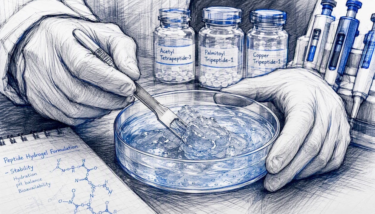
Peptide-loaded hydrogels. Hydrogels extend residence time and can be tuned through crosslink density to control release kinetics, slower crosslinking generally means slower, more sustained peptide release. CyRL‑QN15 loaded onto a zinc-alginate hydrogel nanoparticle system showed improved epithelialization and angiogenesis compared to the free peptide alone, a direct demonstration that formulation choice changed the outcome independent of the peptide’s inherent activity.
Alginate and zinc-alginate dressings. These combine a moisture-retentive matrix with the added benefit of zinc, which has its own supportive role in epithelial repair, offering a practical middle ground between simple gels and engineered nanoparticle systems.
Electrospun nanofibers. These mimic the fibrillar architecture of native ECM, giving cells a structural scaffold to migrate across while also serving as a peptide reservoir. The high surface area can improve both cell adhesion and controlled peptide release, though manufacturing consistency at scale remains a real hurdle.
Peptide-loaded nanoparticles. Encapsulation inside lipid or polymer nanoparticles offers the strongest protease shielding of the topical options, at the cost of the most complex manufacturing process and the highest per-unit cost.
A few formulation variables determine whether any of these systems will actually work in practice:
- Crosslink density controls release rate; too fast wastes the payload, too slow starves the wound of active peptide when it’s needed most.
- Co-delivery of protease inhibitors or ROS scavengers alongside the peptide can meaningfully extend its functional half-life inside the wound.
- Local peptide concentration at the wound interface, not the loaded dose, is the number that actually predicts biological effect.
- Release curves measured in simulated wound fluid correlate far better with in vivo performance than release curves measured in plain saline.
Pro Tip: Run your protease stability and release kinetics testing in simulated wound fluid, not phosphate-buffered saline, before you commit to an animal study. A peptide that looks stable in PBS can fall apart within an hour once it meets the elastase and matrix metalloproteinase activity in real wound exudate, and finding that out after the animal work is an expensive way to learn it.
What Evidence and Models Best Predict Translational Success?
Most peptide evidence for wound healing sits at the in vitro and small-animal in vivo level, and effect sizes tend to look impressive in standardized assays while varying considerably once you move to a different animal model or wound type. That gap is the single most important thing to understand before designing a study around any of these peptides.
In vitro assays form the first screening layer. The scratch (or wound-healing) assay measures keratinocyte or fibroblast migration into a cell-free gap and remains the fastest, cheapest way to get an initial migration readout. Proliferation assays like MTT or BrdU incorporation, alongside Ki‑67 immunostaining, quantify whether cells are actually dividing rather than just moving. Collagen deposition assays (often hydroxyproline quantification or picrosirius red staining) measure matrix output directly. Angiogenesis gets tested through HUVEC tube formation assays, and macrophage cytokine readouts, particularly TGF‑β and IL‑6, indicate immunomodulatory activity. None of this matters if the peptide fails hemolysis or cytotoxicity screening, which should happen in parallel, not as an afterthought.
In vivo models carry more translational weight but also more variability. Full-thickness excisional wound models in rodents remain the standard starting point. Diabetic models, either genetically obese db/db mice or streptozotocin-induced diabetic rodents, are far more clinically relevant given how much human chronic wound burden involves diabetes, yet they’re underused relative to standard models simply because they’re harder and slower to run. Ischemia-reperfusion models add another layer of clinical realism for peritraumatic and pressure wounds. Endpoints across these models typically include re-epithelialization rate, percent wound closure over time, histological assessment of collagen deposition and granulation tissue, and biomechanical tensile strength of the healed tissue.
| Assay or model | What it shows | Key limitation |
|---|---|---|
| Scratch assay | Keratinocyte/fibroblast migration | Doesn’t capture matrix remodeling or angiogenesis |
| Proliferation assay (MTT/BrdU/Ki‑67) | Cell division rate | Doesn’t confirm functional tissue repair |
| HUVEC tube formation | Angiogenic potential | In vitro only, doesn’t reflect in vivo vascular perfusion |
| Full-thickness excisional model | Overall wound closure kinetics | Healthy rodents heal fast; poor diabetic wound proxy |
| Diabetic (db/db or STZ) model | Delayed-healing translational relevance | Slower, more expensive, less standardized across labs |
A few representative case studies show how this plays out in practice. LL‑37 and WHP1 both show dose-dependent migration in HaCaT scratch assays, with WHP1’s peak effect at 62.5 µg/ml alongside macrophage TGF‑β induction. CyRL‑QN15 and Tiger17 have both been tested in diabetic mouse models, where the combination of epithelialization and collagen deposition outcomes suggests activity beyond a single mechanism. A7‑1 stands out for its in vivo angiogenesis data in the C57BL/6 model, showing roughly a 17 percent improvement in wound closure alongside earlier vascularization on angiography. Collagen mimetic peptide carriers, meanwhile, are typically evaluated for structural integration into native collagen rather than for the migration or proliferation endpoints that dominate the other categories.
What Stands Between These Peptides and the Clinic?
Peptides carry strong preclinical promise for wound repair, but the same set of translational bottlenecks show up across nearly every candidate: proteolytic degradation, short functional half-life, formulation complexity, immunogenicity risk, and a regulatory pathway that has no standardized playbook for this drug class yet.
Stability and pharmacokinetics. Wound fluid is a protease-rich environment by design, part of the body’s own debridement process, which is exactly what makes it hostile to unprotected peptides. Linear peptides without stabilizing chemistry often degrade within a narrow window after application, which is why sustained local delivery generally outperforms systemic administration for wound-specific applications. Systemic dosing also raises the exposure risk for a peptide whose bioactivity was optimized for a topical, localized context.
Safety screening. Hemolysis testing against red blood cells, cytotoxicity assays on keratinocytes and fibroblasts, and immunogenicity screens all need to happen early, not after a promising efficacy signal has already generated momentum toward animal studies. Manufacturing introduces its own safety variable: peptide aggregation and synthesis impurities can trigger immune responses unrelated to the peptide’s intended mechanism.
Manufacturability and scale. Solid-phase peptide synthesis works reliably for straightforward linear sequences, but cyclization steps, whether through head-to-tail cyclization, disulfide bridging, or stapling, add purification complexity and cost that scale poorly from milligram research batches to anything resembling clinical-grade production volumes.
Regulatory and endpoint standardization. This may be the least discussed barrier and arguably the most consequential. There’s no widely agreed set of biomarkers or clinical endpoints specific to peptide-based wound therapeutics the way there is for, say, antibiotics or anticoagulants. That absence makes it harder for any single research group’s data to be comparable to another’s, which in turn slows the kind of meta-analysis that regulators eventually want to see before approval.
Pro Tip: If your peptide shows a strong efficacy signal in early screening, don’t wait to run hemolysis and protease-stability testing “later.” Build a basic GLP-compatible toxicology plan alongside your efficacy work from the start. Candidates that show promise but skip early safety characterization tend to stall for months once a funder or reviewer finally asks for the data.
How Should Researchers Design and Test New Wound Peptides?
Template-assisted rational design and deliberate scaffold engineering measurably increase the odds of producing a peptide that’s both potent and non-toxic, and WHP1 is the clearest illustration of the approach working as intended.
A practical preclinical roadmap generally follows six stages:
- Target selection and mechanism mapping. Decide which wound phase you’re targeting, inflammatory, proliferative, or remodeling, before you design anything. A peptide built for angiogenesis will underperform if deployed against an early hyper-inflammatory bottleneck.
- In silico design and motif selection. Sequence alignment against known active peptides and computational stability prediction narrow the candidate pool before a single molecule gets synthesized.
- Synthesis and initial in vitro screens. Protease stability, hemolysis, and scratch or proliferation assays should run together, not sequentially, so a toxicity red flag surfaces before you’ve invested in downstream work.
- Formulation and release testing. Simulated wound fluid testing, not saline, determines whether your delivery system will actually protect the peptide once it’s inside a real wound.
- Small-animal proof of concept. Full-thickness and diabetic wound models, ideally both, with a genuine dose-response curve rather than a single dose comparison against control.
- GLP-compatible safety readouts and IND-enabling studies. This is where most academic pipelines stall, not for lack of efficacy data but for lack of the standardized toxicology package regulators expect.
WHP1’s own development followed a version of this logic: template-assisted design led to a candidate that showed migration, proliferation, antioxidant protection, and macrophage TGF‑β induction together in a single phenotypic screening panel, an approach that let researchers de-risk the candidate on multiple mechanistic fronts before ever reaching for an animal model.
For reproducibility, and frankly for getting published in a journal worth publishing in, a peptide wound-healing study should report: the exact sequence and purity (with a Certificate of Analysis), the dose range tested, the formulation and delivery vehicle used, full animal model details including strain and comorbidity status, and the specific endpoints measured with their assay methodology. Papers that skip any of these make replication, and honest comparison against other candidates, essentially impossible.
Where Does Peptide Therapy for Wound Healing Stand Today?
Most of the strongest peptide evidence for wound repair remains preclinical. A small number of cyclic and synthetic scaffolds have advanced into early translational development, but large randomized controlled trials specific to peptide-based wound therapeutics are still uncommon relative to the volume of preclinical publications.
The near-term research priorities that would actually move this field forward include:
- Standardized pharmacokinetic and pharmacodynamic characterization specifically in wound fluid, not serum or plasma, since the degradation environment is fundamentally different
- Head-to-head formulation comparisons, since delivery system choice currently varies so much between studies that cross-study comparison is close to meaningless
- Combination therapy studies pairing peptides with advanced dressings rather than testing peptides in isolation
- Biomarker standardization across TGF‑β, VEGF, and MMP ratios, so different labs are measuring outcomes on comparable terms
- Deeper representation of diabetic and ischemic wound heterogeneity, since healthy-animal healing kinetics predict clinical performance in comorbid patients poorly
Trial design features likely to matter most to regulators evaluating this drug class include objective, imaging-based wound area metrics rather than clinician estimation, time-to-re-epithelialization as a primary endpoint, histologic ECM assessment as a secondary endpoint, and a safety panel that specifically accounts for systemic peptide exposure even in topically applied products.
Looking ahead, the most plausible near-term advances are modular cyclic scaffolds that can be adapted to different wound phases by swapping functional motifs, micropeptide therapeutics derived from long noncoding RNA smORFs (a genuinely new peptide discovery pipeline rather than an incremental one), and multifunctional dressings that combine a peptide payload with a protease inhibitor or antioxidant co-therapy in a single delivery matrix.
Where should researchers entering this field put their effort first?
Prioritize mechanism-matched peptides over broadly “promising” ones. A peptide with excellent migration data but no plan for surviving wound fluid protease activity is a paper, not a candidate. The order that matters, in my view: first, solve local delivery and protease protection, because no mechanism matters if the peptide degrades before it acts. Second, choose animal models that reflect actual clinical comorbidity, diabetic and ischemic models, not just healthy young rodents that heal on their own within days regardless of intervention. Third, insist on standardized reporting and full Certificate of Analysis transparency for every peptide batch used in a study, since batch-to-batch variability quietly undermines more wound-healing papers than anyone likes to admit.
My honest forecast: clinical impact will arrive first through topical dressings built around rationally designed cyclic scaffolds combined with multifunctional hydrogels, not through systemic peptide therapy. That’s where the delivery problem is most tractable and where the safety margin is widest.
A note on sourcing lab-grade peptides for this research
Every mechanism and model discussed above depends on one unglamorous variable that determines whether your data means anything: peptide purity and batch consistency. Data Peptides supplies lab-grade research peptides, including compounds relevant to tissue repair research such as BPC‑157 and TB‑500 and LL‑37, each backed by a Certificate of Analysis documenting purity for exactly the kind of reproducibility your methods section needs. All products are sold strictly for laboratory research use, not for human or animal consumption, and researchers remain responsible for following their institution’s own regulatory and ethical protocols for any in vivo work. If your next experiment needs a reliably sourced reagent rather than another variable to control for, the shop page lists current research peptide inventory with COAs attached to each batch.
Sources
- Rational engineering unlocks the therapeutic potential of WHP1: A revolutionary peptide poised to advance wound healing | PLOS One
- More than skin deep: cyclic peptides as wound healing and regenerative agents (Frontiers in Cell and Developmental Biology)
- NCBI PMC article (PMC9388858)
- Wound healing induced by new synthetic peptide, A7-1, in C57BL/6 mouse model | BioMedical Engineering OnLine
This article is general information, not a substitute for advice from a qualified doctor. Consult a qualified healthcare professional about your own circumstances before acting on anything here.
FAQ
What Are the Best Peptides for Wound Healing Research?
Cyclic scaffolds like CyRL‑QN15 and rationally designed synthetics like WHP1 currently carry the strongest combination of mechanistic evidence and protease stability, though LL‑37 remains the most-studied natural antimicrobial option.
How Do Peptides Aid Healing Compared to Growth Factors?
Peptides often engage the same receptor and signaling pathways as growth factors (VEGF, TGF‑β induction) but at a fraction of the manufacturing cost and with better tunability for protease resistance through cyclization.
Are Collagen Peptides for Wounds Different From Signaling Peptides?
Yes. Collagen mimetic peptides physically integrate into damaged collagen fibrils to reinforce matrix structure, while signaling peptides like LL‑37 or WHP1 act through receptor engagement to trigger migration, proliferation, or cytokine release.
Is Peptide Therapy for Injury Recovery Ready for Clinical Use?
Most peptide wound-healing evidence remains preclinical. A few cyclic and synthetic candidates have entered early translational development, but large clinical trials specific to this drug class are still limited.
Where Can Researchers Source Lab-Grade Peptides for These Studies?
Data Peptides supplies research-use peptides relevant to tissue repair studies, including BPC‑157, TB‑500, and LL‑37, each with a Certificate of Analysis for batch verification.