
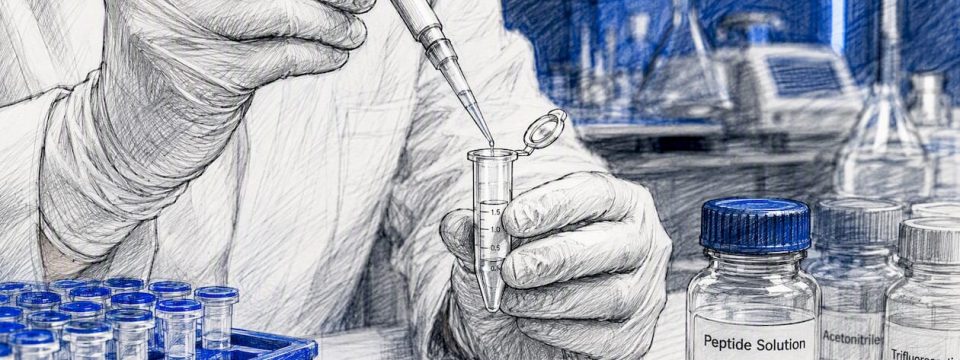
Anti-inflammatory peptides work by interrupting the same signaling cascades that drive chronic disease: NF-κB activation, MAPK phosphorylation, and oxidative stress accumulation. Some, like the tight-junction-inducing fragments of alpha-1 antitrypsin, do something different entirely, physically rebuilding the barriers that inflamed tissue loses. The honest caveat: most of this evidence comes from cell cultures and rodent models, not human trials.
That gap matters more than most sourcing pages admit. A Frontiers in Immunology review documents bioactive peptides suppressing NF-κB and MAPK signaling across gut, skin, lung, and joint models, with reduced oxidative stress as a consistent secondary effect. BPC-157, one of the most cited research peptides in this space, has a deep rodent literature for gut and tendon repair but no published Phase II or III human trials backing those endpoints. Structurally, the peptides that tend to work share a pattern: small size, under 1 kDa and 2 to 20 amino acids, with a mix of hydrophobic and positively charged residues that lets them cross cell membranes and reach intracellular targets.
Pro Tip: When you’re scanning a peptide’s literature, check whether the anti-inflammatory claim comes from a serum cytokine panel, a histology score, or an actual clinical outcome measure. Those are three very different confidence levels, and abstracts rarely make the distinction obvious.
Key Takeaways
Anti-inflammatory peptides work primarily by suppressing NF-κB and MAPK signaling or by physically restoring tissue barriers, but almost all of this evidence remains preclinical rather than clinically validated.
| Point | Details |
|---|---|
| Match mechanism to tissue | Choose gut-targeted peptides like KPV for mucosal inflammation and barrier-repair peptides like A1AT JIPs for epithelial damage models. |
| Verify purity independently | Require HPLC purity above 98%, mass spectrometry confirmation, and a batch-specific Certificate of Analysis before use. |
| Prioritize PK/PD early | Characterize pharmacokinetics and pharmacodynamics before designing efficacy studies to avoid dosing and delivery mismatches. |
| Read evidence levels honestly | Treat rodent colitis or tendon data as preclinical signal, not a substitute for phase II or III human trial evidence. |
| Source from a verified supplier | Data Peptides provides third-party tested, research-use-only peptides with full COAs for reproducible study design. |
Table of Contents
- What Are Anti-Inflammatory Peptides?
- How Do Peptides Reduce Inflammation at the Molecular Level?
- Which Peptides Are Best Studied for Inflammation?
- Where Do Anti-Inflammatory Peptides Show the Strongest Results?
- Why Do Peptides Struggle to Reach the Clinic?
- Is There Clinical Evidence for Peptide Therapy for Pain and Inflammation?
- How Should Researchers Source and Verify Peptides for Lab Work?
- Where Is Peptide-Based Inflammation Research Headed?
- Sourcing Research-Grade Peptides for Inflammation Studies
- Sources
- FAQ
What Are Anti-Inflammatory Peptides?
Anti-inflammatory peptides, sometimes called bioactive peptides (BAPs) in the food-science literature, are short chains of amino acids that modulate immune signaling rather than blocking a single enzyme the way a classical NSAID does. Where ibuprofen inhibits cyclooxygenase directly, a peptide like KPV works by entering an intestinal epithelial cell and preventing NF-κB from moving into the nucleus in the first place. That upstream action is what makes peptides interesting to researchers: one molecule can touch several downstream pathways instead of one narrow target.
The field splits into four rough categories, and the distinction matters when you’re deciding what to study or source.
- Endogenous immunoregulatory peptides — sequences the body already produces or derives from its own proteins, such as Ac-SDKP, a natural regulator of tissue fibrosis and inflammation.
- Food-derived bioactive peptides — fragments released during digestion of milk casein, fish protein, or plant sources, many showing anti-inflammatory activity in cell and animal models.
- Antimicrobial peptides with immunomodulatory roles — LL-37 is the best-known example, a human cathelicidin that fights pathogens while also shaping cytokine release and wound repair.
- Synthetic peptides and peptidomimetics — engineered sequences like BPC-157 and TB-500 designed or discovered specifically for stability and targeted activity, plus peptidomimetic variants built to resist enzymatic breakdown.
Structurally, the peptides that show reliable anti-inflammatory activity share physical traits. According to a Food & Function review, an estimated 77% of bioactive peptides identified in the literature come from what researchers call alternative or sustainable protein sources, insects like Tenebrio molitor, agro-industrial byproducts, sunflower protein, and similar unconventional feedstocks rather than traditional animal tissue. Hydrophobic residues (valine, isoleucine, proline) and positively charged residues (histidine, arginine, lysine) show up disproportionately often in active sequences, likely because that combination helps peptides cross intestinal and cell membranes.
Anti-inflammatory peptides (also called anti-inflammatory bioactive peptides) are short amino acid chains, generally under 20 residues, that modulate immune cell signaling and cytokine release rather than inhibiting a single inflammatory enzyme.
How Do Peptides Reduce Inflammation at the Molecular Level?
Three pathways dominate the mechanistic literature: NF-κB, MAPK, and JAK-STAT. Most anti-inflammatory peptides hit at least one of these, and the strongest candidates seem to touch two or more simultaneously.
- NF-κB inhibition — many peptides block nuclear translocation of NF-κB, the transcription factor responsible for switching on genes for TNF-α, IL-6, and IL-1β. KPV, a tripeptide derived from alpha-MSH, is the clearest example, shown to inhibit NF-κB nuclear translocation in intestinal epithelial cells and reduce colitis severity in rodent models.
- MAPK modulation — the Frontiers in Immunology review documents peptides that dampen p38 and ERK phosphorylation, reducing downstream production of inflammatory mediators in skin, gut, and lung tissue models.
- JAK-STAT effects — less studied than NF-κB but increasingly implicated in peptide research targeting chronic inflammatory states where cytokine receptor signaling stays switched on.
- Oxidative stress reduction — several peptides, including casein-derived fragments and antler-derived sequences, show direct antioxidant activity, scavenging reactive oxygen species that otherwise amplify the inflammatory cascade.
- Cytokine release modulation — beyond transcription factor inhibition, some peptides act more directly on cytokine secretion or receptor binding, shifting the overall inflammatory tone of a tissue.
At the cellular level, the effects show up as macrophage polarization from the pro-inflammatory M1 phenotype toward the reparative M2 phenotype, reduced neutrophil infiltration into damaged tissue, and, in the case of barrier peptides, direct induction of tight-junction proteins between epithelial cells. That last mechanism is where the field has made one of its more interesting recent discoveries. C-terminal fragments of alpha-1 antitrypsin, known as tight-junction-inducing peptides or JIPs, insert into epithelial membranes, activate G13 signaling, and physically assemble claudin proteins into functional tight junctions. In DSS-induced colitis mouse models, synthetic JIP peptides restored barrier integrity and prevented colitis from developing, and human homologs of the same peptide (JIPh42, also called CAAP48) showed comparable tight-junction activity in vitro.
Pro Tip: A peptide that suppresses cytokine release in a 96-well plate assay is not the same as a peptide that will do the same thing in a living organism with intact clearance mechanisms and an active immune system. Preclinical pathway data tells you the peptide can theoretically hit a target; it does not tell you the dose, route, or half-life needed to hit that target reliably in a mammal.
Which Peptides Are Best Studied for Inflammation?
Seven sequences account for most of the citable evidence in this space, and they sit at very different points on the evidence spectrum. Some have decades of mechanistic study behind them; others are recent discoveries still working out their basic pharmacology.
BPC-157, a 15-amino-acid fragment derived from a protective protein found in gastric juice, has the broadest rodent evidence base of any peptide on this list. Studies link it to NF-κB suppression, nitric oxide system modulation, and accelerated tissue repair across gut, tendon, and even systemic injury models. No phase II or III human trials have been published for these endpoints, which makes BPC-157 a case study in the gap between rodent promise and clinical proof.
KPV (Lys-Pro-Val) is a tripeptide fragment of alpha-MSH with the strongest gut-specific translational signal in the group. It gets absorbed by intestinal epithelial cells and directly blocks NF-κB nuclear translocation, cutting TNF-α and IL-6 output in colitis models. Because it acts locally in the gut rather than requiring systemic circulation, KPV is one of the more plausible near-term candidates for oral formulation work.
TB-500, a fragment of thymosin beta-4, works through a different mechanism entirely: it regulates actin dynamics, the cellular scaffolding that controls cell migration. That gives it a role in reducing inflammatory cell infiltration while promoting the kind of directed cell movement tissue needs to repair itself, which is why it shows up frequently in tendon and wound-healing research.
Ac-SDKP (N-acetyl-seryl-aspartyl-lysyl-proline) is an endogenous tetrapeptide the body produces naturally as a regulator of tissue fibrosis and inflammatory cell recruitment. Its natural origin gives it a different risk profile than fully synthetic peptides, though human clinical data remains limited.
Antiflammin, derived from a region of the annexin A1 protein, was among the earlier anti-inflammatory peptides characterized in the literature. A broad review of anti-inflammatory peptide mechanisms documents its immunomodulatory activity alongside other natural sequences like bee venom fragments and bovine casein derivatives.
LL-37, the only peptide on this list that’s also a well-characterized antimicrobial agent, shapes cytokine release and wound-healing signaling in addition to its pathogen-fighting role, making it a useful example of how immune defense and inflammation regulation overlap in a single sequence.
A1AT C-terminal peptides (JIPs/CAAP48) stand apart from the rest of the list because they work through tight-junction assembly rather than cytokine suppression, which is arguably the field’s most conceptually distinct mechanism, repairing the barrier itself instead of just quieting the immune response.
| Peptide | Primary mechanism | Typical model | Highest human signal |
|---|---|---|---|
| BPC-157 | NF-κB suppression, nitric oxide modulation | Rodent gut, tendon, systemic injury | Preclinical only |
| KPV | NF-κB nuclear translocation blockade | Murine DSS colitis | Preclinical only |
| TB-500 | Actin dynamics, cell migration | Rodent and equine tendon/wound models | Preclinical only |
| Ac-SDKP | Fibrosis and cytokine recruitment regulation | Rodent fibrosis and cardiac injury models | Biomarker studies in humans |
| Antiflammin | Annexin A1-derived immunomodulation | Classical rodent inflammation models | Preclinical only |
| LL-37 | Antimicrobial plus cytokine/wound healing signaling | Human keratinocyte and rodent skin models | Early clinical/biomarker data |
| A1AT JIPs (CAAP48) | Tight-junction assembly via G13 activation | Murine colitis, human epithelial cell lines | In vitro human tissue data |
The pattern across this table is stark: every sequence has a solid preclinical foundation, and almost none has cleared a phase II or III human trial for inflammation-specific endpoints. That’s not a reason to dismiss the mechanisms, rodent-to-human extrapolation has a real track record in pharmacology generally, but it is a reason to read “clinically proven” claims about any of these peptides with real skepticism. LL-37 and Ac-SDKP have the most human-adjacent data because both occur naturally in human tissue and have been measured as biomarkers in patient samples, which is a different evidentiary category than an interventional trial.
Where Do Anti-Inflammatory Peptides Show the Strongest Results?
Organ-specific evidence tends to cluster around a few systems where researchers have built up repeated model data, and the strength of that evidence varies considerably by tissue.
Gut and inflammatory bowel disease models have the deepest bench of peptide research, largely because DSS-induced colitis in mice is a well-standardized, reproducible model. KPV and BPC-157 both show reduced TNF-α and IL-6 alongside improved histology scores in colitis models, and the A1AT-derived JIPs go a step further by preventing colitis outright through tight-junction restoration rather than just dampening cytokine output.

Skin and wound healing research features LL-37 prominently, given its dual role as an antimicrobial and immune signaling peptide in keratinocyte models, alongside TB-500 for its actin-mediated effects on cell migration during repair.
Lung inflammation models, covering both pneumonia-type injury and allergic inflammation, show peptide-mediated reductions in inflammatory mediator release, though this application area has fewer named, widely-cited peptide candidates compared to gut and skin research.
Joint and tendon applications rely heavily on TB-500 and BPC-157 data, including equine tendon studies that provided some of the earliest large-animal evidence for these sequences outside rodent work.
Ocular inflammation research is the thinnest of the five systems in terms of named peptide candidates, though the same NF-κB and oxidative-stress mechanisms documented elsewhere are beginning to show up in early ocular surface studies.
| Model | Intervention | Primary outcome | Evidence level |
|---|---|---|---|
| Murine DSS colitis | KPV (oral/local) | Reduced TNF-α and IL-6, improved histology | Preclinical |
| Murine DSS colitis | A1AT JIP peptides | Restored tight junctions, colitis prevented | Preclinical, human tissue data supportive |
| Rodent/equine tendon injury | TB-500 | Reduced inflammatory infiltration, faster repair | Preclinical |
| Rodent gastric/tendon injury | BPC-157 | Reduced pro-inflammatory cytokines, faster repair | Preclinical |
| Human keratinocyte/rodent skin | LL-37 | Modulated cytokine release, wound healing support | Early clinical/biomarker |
The gut is the clearest case where peptides act locally rather than needing full systemic distribution, which is exactly why oral delivery research for tripeptides like KPV has gotten so much attention: the target tissue is right there in the gastrointestinal tract, and the peptide doesn’t need to survive circulation to reach it. Joint and tendon applications are the opposite case, usually requiring injection or some form of systemic or local-depot delivery to reach a target that isn’t accessible from the gut lumen.

Why Do Peptides Struggle to Reach the Clinic?
Enzymatic degradation is the single biggest reason a promising peptide never makes it past animal models. Gastrointestinal and serum proteases chew through unmodified peptide bonds within minutes in many cases, which is why oral bioavailability is such a persistent problem for this entire drug class. Add rapid renal clearance, most small peptides get filtered out of circulation quickly, and limited membrane permeability, since many sequences can’t cross a cell membrane without help, and you have the three barriers that define peptide pharmacology.
Researchers have developed a handful of mitigation strategies that show up repeatedly across the literature.
- Nanoparticle encapsulation protects a peptide from enzymatic attack until it reaches its target tissue; encapsulated KPV formulations have shown improved outcomes in murine colitis models compared to unencapsulated delivery.
- PEGylation attaches polyethylene glycol chains to a peptide, extending its half-life in circulation by reducing renal clearance and shielding it from some protease activity.
- Peptidomimetic design replaces vulnerable peptide bonds with non-natural linkages that proteases can’t recognize, trading some of the natural sequence’s flexibility for stability.
- Local and oral delivery routes work best for gut-targeted peptides like KPV precisely because the target tissue sits in the delivery path, sidestepping the need for systemic survival entirely.
The trade-offs are real. PEGylation extends half-life but can reduce a peptide’s binding affinity or cell penetration. Nanoparticle encapsulation improves stability but adds manufacturing complexity and cost. Peptidomimetics solve the protease problem but require redesigning the molecule almost from scratch, which means starting the preclinical evidence chain over again.
Pro Tip: If you’re designing a translational study, run pharmacokinetic and pharmacodynamic characterization before you invest in efficacy endpoints. A peptide that clears from serum in eight minutes needs a completely different dosing strategy than one with a six-hour half-life, and that distinction should shape your protocol before you touch an outcome measure.
Is There Clinical Evidence for Peptide Therapy for Pain and Inflammation?
Clinical evidence remains thin across almost every peptide covered here, and researchers considering peptide therapy for pain or inflammation should treat that as the starting assumption, not a footnote. KPV has the strongest gut-specific translational signal of the group, based on consistent effects across multiple rodent colitis models, but that still falls short of a published phase II or III human trial. BPC-157’s rodent data is broad and repeated across labs, yet the same absence of controlled human trials applies.
Experts in the field consistently flag translation as the primary bottleneck, not the underlying biology. Peptides tend to show high target selectivity compared to broad-acting anti-inflammatories like corticosteroids, which is a real theoretical advantage, but selectivity in a mouse model doesn’t guarantee an equivalent effect or safety profile in humans. That’s the gap researchers are still working to close.
A practical safety checklist for anyone designing early-stage human or advanced preclinical work:
- Immunogenicity risk — any peptide, especially synthetic or modified sequences, carries a theoretical risk of triggering an antibody response that could neutralize its activity or cause adverse reactions.
- Wound-healing interference — peptides that suppress inflammation too broadly could theoretically impair the early inflammatory phase that normal wound repair depends on, a consideration for study designs involving injury models.
- Theoretical tumor-promoting concerns — some growth-factor-adjacent peptides warrant careful monitoring in longer-duration studies, given that unchecked cell proliferation signaling is a legitimate research question, not settled fact in either direction.
- Storage and handling safety — lyophilized peptides and reconstituted solutions have specific stability windows, and lab handling protocols should account for degradation risk affecting both results and reagent safety.
- Endpoint selection — translational trials need endpoints that map cleanly from the animal model to a human-relevant outcome; a cytokine panel that moved in a mouse doesn’t automatically translate to a validated human biomarker.
None of this diminishes the legitimacy of the mechanistic case for these peptides. It does mean that “proven” is the wrong word for where most of this evidence currently sits, and researchers designing new studies should plan for that gap rather than around it. This article and the products referenced here are intended strictly for laboratory research use, not for human or animal treatment, and nothing here should be read as medical advice.
How Should Researchers Source and Verify Peptides for Lab Work?
Sourcing quality is where a lot of otherwise-solid research falls apart, and the checklist here is short but non-negotiable for reproducible work.
- HPLC purity of 98% or higher, confirmed by high-performance liquid chromatography, is the baseline standard for any peptide entering a serious research protocol.
- Mass spectrometry confirmation verifies that the sequence you ordered is the sequence you received, catching synthesis errors that purity percentage alone won’t reveal.
- Endotoxin testing (LAL assay) matters enormously for any in vivo or cell-culture work, since endotoxin contamination can independently trigger inflammatory responses and confound your entire dataset.
- Certificate of Analysis (COA) availability for every batch, not just a generic product-level document, so you can trace exactly which lot went into which experiment.
- Storage protocols: lyophilized peptides typically hold stability at −20°C for extended periods, while reconstituted solutions generally need 2–8°C storage and a defined use window before degradation becomes a concern.
- Chain-of-custody documentation that lets you reconstruct exactly how a peptide was handled from manufacture to your bench, essential for any study aiming at reproducibility.
Beyond sourcing, a handful of study-design habits separate reproducible peptide research from noisy, unreplicable results. Confirm sequence identity independently by LC-MS rather than trusting a vendor’s label alone. Run stability testing under your own intended storage and use conditions rather than assuming published stability data transfers exactly. Build pharmacokinetic and pharmacodynamic assays into early study phases, not as an afterthought once efficacy signals look promising. Include immunogenicity assays where the study duration or dosing frequency makes that risk plausible. And always run appropriate vehicle and negative controls, peptide research is particularly vulnerable to batch effects that a poorly controlled study will misattribute to biological activity.
Pro Tip: Batch-to-batch variability is one of the most underestimated sources of failed replication in peptide research. Batch-match your reagents across an entire experimental series when possible, and request independent third-party testing results, not just the vendor’s in-house COA, for any peptide going into a study you intend to publish.
Data Peptides’ laboratory standards and best-practices resource covers these handling and documentation requirements in more depth for researchers building out a new protocol.
Where Is Peptide-Based Inflammation Research Headed?
The field’s near-term progress is going to come from two directions: better barrier-repair peptides built on the tight-junction model that A1AT-derived JIPs pioneered, and better oral formulations for gut-targeted tripeptides like KPV, where nanoparticle encapsulation has already shown it can meaningfully improve bioavailability in preclinical work. Both of those are incremental, unglamorous advances, but they’re the kind that actually move a peptide closer to a human trial rather than just adding another mouse study to an already crowded literature.
The bottleneck isn’t imagination. Researchers have identified dozens of mechanistically interesting sequences, and new ones surface in the literature regularly. The bottleneck is rigorous human trial infrastructure, and that’s a slower, more expensive problem than finding the next candidate peptide. If there’s one thing the field should prioritize over discovering novel sequences, it’s mechanism-to-target matching done early and pharmacokinetic and pharmacodynamic characterization done before efficacy claims get made. A peptide that works beautifully on the wrong tissue, or at the wrong dose, or with the wrong half-life, will look like a failed drug when it was actually just a mismatched experiment.
Data Peptides exists to support that work with material researchers can actually trust: peptides where the purity claim matches the certificate, and the certificate matches the batch. That’s a smaller contribution than solving the translation problem outright, but it’s the one a peptide supplier can actually make, and it matters more than most researchers realize until they’ve been burned by a batch that didn’t match its label.
Sourcing Research-Grade Peptides for Inflammation Studies
Every peptide covered in this article, BPC-157, KPV, TB-500, and the rest, is only as useful to your research as the purity and documentation behind the batch you’re working with.
For gut-inflammation and tissue-repair research specifically, the peptide 157 research overview covers sourcing details relevant to BPC-157 study design, and Data Peptides supports bulk ordering for multi-batch studies along with storage and handling guidance and direct technical support for researchers structuring PK/PD or efficacy protocols. Every product is sold strictly for laboratory research use, not for human or animal consumption.
If you’re planning your next inflammation study and need a peptide source you can document with confidence, browse Data Peptides’ shop to see current lab-grade inventory and Certificates of Analysis before you place your next order.
Sources
- Peptides and protein hydrolysates exhibiting anti-inflammatory activity: sources, structural features and modulation mechanisms – Food & Function (RSC Publishing)
- Frontiers in Immunology review: therapeutic effects of bioactive peptides on inflammation (review article)
- Science Advances: TJ-inducing peptides from A1AT restore epithelial barrier and prevent colitis (JIP study)
- PubMed: review on anti‑inflammatory peptides and their mechanisms (overview article)
FAQ
What is the strongest supplement for inflammation?
No single peptide has clinical trial evidence strong enough to call it “the strongest.” BPC-157 and KPV have the broadest preclinical evidence bases, but both remain backed primarily by rodent studies rather than published human trials.
How do peptides reduce inflammation differently than NSAIDs?
Peptides typically act upstream, blocking transcription factors like NF-κB before inflammatory genes get switched on, while NSAIDs inhibit a specific downstream enzyme like cyclooxygenase. That upstream action lets a single peptide influence several inflammatory pathways at once rather than one narrow target.
Are anti-inflammatory peptides safe for human use?
Most anti-inflammatory peptides discussed in the research literature have only been tested in preclinical models, so human safety data is limited, and theoretical risks like immunogenicity haven’t been fully characterized. This information is general and not a substitute for professional medical guidance; these peptides are sold strictly for laboratory research use, not for human treatment.
Which peptide is best for gut inflammation specifically?
KPV has the strongest gut-specific translational signal because it acts locally on intestinal epithelial cells and blocks NF-κB nuclear translocation directly, with nanoparticle-encapsulated formulations showing improved outcomes in murine colitis models.
Why do peptides work in animal studies but not always in humans?
Enzymatic degradation, rapid renal clearance, and limited membrane permeability all reduce a peptide’s effective dose and duration in circulation, and these pharmacokinetic barriers often behave differently across species. That mismatch is a major reason rodent efficacy doesn’t automatically predict human clinical results.