

IGF-1 LR3 is a long-Arg3 IGF-1 analogue engineered for low IGF-binding protein affinity, which makes its receptor signaling and apparent potency highly context-dependent. That single property is the reason identical doses produce different outcomes across cell lines, species, and physiological states, and why researchers designing or interpreting LR3 experiments need to account for IGFBP status and systemic endocrine shifts before drawing conclusions.
TL;DR:
- LR3’s receptor signaling varies significantly depending on IGFBP presence, causing potency to invert between IGFBP-secreting and non-secreting systems.
- Continuous LR3 infusion in animals often suppresses growth hormone and IGFBP-3 levels, leading to negative or neutral effects on growth and metabolism.
- Accurate measurement of LR3 requires an specific ELISA targeting its Arg3 epitope, with strict quality control, careful sample handling, and reporting of IGFBP status.
- Systemic endocrine feedback, particularly GH suppression and amino acid decreases, should be primary safety markers monitored in LR3 studies.
- LR3 shows potential for muscle research but has failed to demonstrate growth benefits in nutrient-limited models, with long-term safety data still lacking.
Table of Contents
- What Is IGF-1 LR3 and Why Does Its Structure Matter?
- What Do the Animal Studies Show About LR3?
- How Should Researchers Measure LR3 Accurately?
- What Systemic Effects Should Researchers Monitor for Safety?
- Where Does LR3 Show Real Therapeutic Promise, and Where Doesn’t It?
- Why Reagent Quality Determines Whether Your LR3 Data Is Reproducible
- What This Research Actually Tells Us About LR3
- Sourcing Research-Grade LR3 for Reproducible Work
- Sources
- FAQ
What Is IGF-1 LR3 and Why Does Its Structure Matter?
The molecule researchers call IGF-1 LR3, or long [Arg3] IGF-1, differs from native IGF-1 in two structural details that change everything about how it behaves in a system. It carries a 13 amino acid extension on the N-terminus and a substitution of arginine for glutamic acid at position 3. Those two changes were characterized in the foundational work by Francis et al. in the Journal of Molecular Endocrinology, which set out to test whether N-terminal extensions and receptor binding could be decoupled from IGFBP binding.
They largely succeeded. The Arg3 substitution disrupts the analogue’s ability to bind IGF-binding proteins (IGFBPs), the circulating and tissue-bound proteins that normally sequester IGF-1 and regulate how much reaches its receptor. Native IGF-1 spends most of its circulating life bound to IGFBP-3 and an acid-labile subunit, tucked away from receptors until release is triggered. LR3 mostly skips that step.
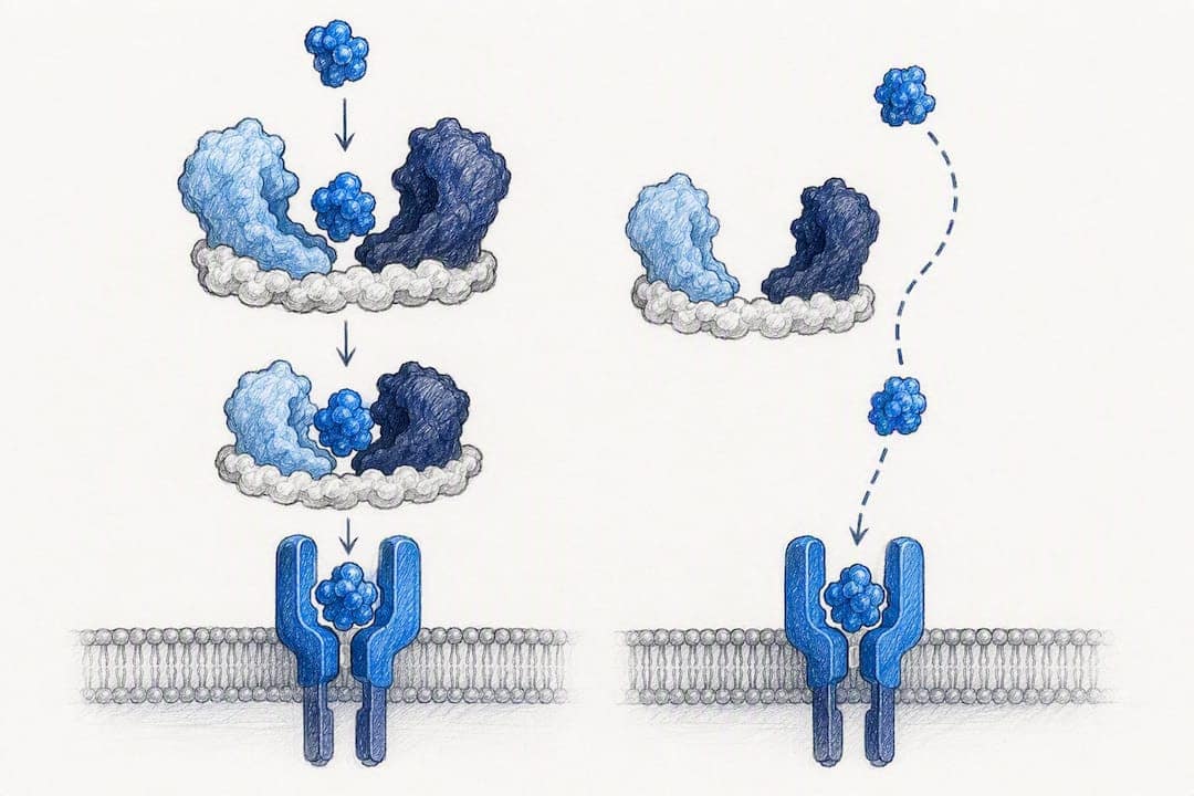
This is the mechanism that defines everything downstream. When IGFBPs are present in the experimental system, less of them can grab hold of LR3, so more of the analogue stays free to engage the IGF-1 receptor. When IGFBPs are absent or minimal, LR3’s altered N-terminus can actually reduce its receptor engagement relative to native IGF-1, because part of the structural extension that blocks IGFBP binding also modestly interferes with receptor fit.
Francis and colleagues demonstrated this directly by comparing potency across multiple cell line types:
- In IGFBP-secreting cell systems, long-Arg3 IGF-1 analogues showed increased relative biological potency compared with native IGF-1, because less analogue was lost to IGFBP sequestration.
- In non-IGFBP-secreting cell systems, the same analogues showed reduced potency relative to native IGF-1, because the receptor-binding advantage from IGFBP evasion had nothing to work against.
- The direction of the potency shift depended entirely on the IGFBP status of the system being tested, not on the analogue’s intrinsic receptor affinity alone.
Statistic callout: The Francis et al. dataset remains the reference point for LR3’s defining behavior: potency inverts direction between IGFBP-secreting and non-secreting systems, which is why a single “fold-change potency” number for LR3 versus native IGF-1 doesn’t exist independent of context. Read the original mechanistic analysis.
This has a direct, practical consequence for how you design and interpret in vitro versus in vivo work. Cell culture media formulations vary widely in serum content and endogenous IGFBP production, meaning two labs running “the same” LR3 dose-response curve on different cell lines can get contradictory potency rankings. A serum-free myoblast assay with minimal IGFBP secretion behaves nothing like a hepatocyte culture that’s actively secreting IGFBP-3.
Plasma is a different animal altogether. Circulating IGFBPs, especially IGFBP-3 in complex with the acid-labile subunit, create a reservoir effect that native IGF-1 depends on for its half-life and distribution. LR3 largely bypasses that reservoir, which changes its pharmacokinetics, its tissue distribution, and its systemic hormonal feedback in ways that don’t show up in a static binding assay. Anyone extrapolating from an isolated cell assay to whole-animal physiology needs to treat IGFBP context as an independent variable, not a footnote. The mechanistic overview of LR3’s research applications covers this sequence-function relationship in more depth for researchers building out protocol documentation.
What Do the Animal Studies Show About LR3?
Two studies anchor most of what’s actually known about LR3 in a living, IGFBP-competent system, and both point toward caution rather than confirmation of a straightforward growth-promoting effect.
The first is a 1997 porcine infusion study that remains the most cited dose-response dataset for LR3 in vivo. Pigs received a 4-day continuous infusion of Long [R3] IGF-1 at a high dose. Instead of the anabolic boost a naive potency argument might predict, the animals showed decreased average daily gain and reduced food intake. Endocrine markers moved in a coordinated direction: plasma IGFBP-3 dropped, endogenous IGF-1 fell, and insulin declined. Co-administering porcine growth hormone alongside the LR3 infusion produced no rescue effect, according to the original pig infusion study.
That result tells you something important: flooding a system with a low-IGFBP-binding IGF-1 receptor agonist doesn’t just amplify local receptor signaling. It triggers negative feedback on the somatotropic axis strong enough to suppress the animal’s own growth hormone and IGF-1 production, and the net effect on whole-body growth was negative, not positive.
The second anchor study is more recent. A 2024 investigation published in the American Journal of Physiology-Endocrinology and Metabolism infused LR3 into growth-restricted fetal sheep at approximately 1.17 μg·kg⁻¹·h⁻¹ continuously for one week, a model designed to test whether boosting IGF receptor signaling could rescue fetal growth restriction (FGR). It didn’t. The fetal-sheep study found no improvement in fetal body weight, insulin, glucose, or oxygen levels, and glucose-stimulated insulin secretion (GSIS) showed no benefit either. Circulating amino acids actually decreased significantly (P = 0.0232).
| Study | Species/model | Dose and duration | Primary outcome |
|---|---|---|---|
| Pig infusion, 1997 | Growing pigs | a high daily dose for a 4-day continuous infusion | Reduced growth rate, food intake, GH (~23% fall), IGFBP-3, endogenous IGF-1, insulin |
| Fetal-sheep infusion, 2024 | Growth-restricted fetal sheep | ~1.17 μg·kg⁻¹·h⁻¹, 1-week continuous infusion | No improvement in weight, insulin, glucose, oxygen, or GSIS; decreased amino acids |
Neither study used LR3 in an oral or bolus format; both relied on continuous infusion, which matters for interpreting dose comparisons across the literature. A bolus injection produces a transient receptor-saturation spike followed by clearance, while continuous infusion sustains a steady-state exposure that better reveals chronic endocrine feedback, which is likely why both studies picked up the GH and IGFBP-3 suppression signal so clearly.
A few translational patterns are worth flagging directly:
- Species-specific endocrine feedback loops mean a dose that’s endocrine-neutral in one model can be strongly suppressive in another; scaling by body weight alone is not sufficient.
- Nutrient-limited models (fetal growth restriction, caloric restriction protocols) appear especially prone to null or negative results, because receptor signaling can’t compensate for a substrate ceiling.
- Both anchor studies used relatively short infusion windows (4 days and 1 week); nothing in the current literature establishes what happens with chronic, multi-week LR3 exposure in a whole-animal model.
How Should Researchers Measure LR3 Accurately?
Getting a clean read on LR3 concentration in plasma or media is harder than it looks, because generic IGF-1 assays weren’t built to tell LR3 apart from native IGF-1 and IGF-2, and IGFBPs can distort results if the assay isn’t designed to handle them.
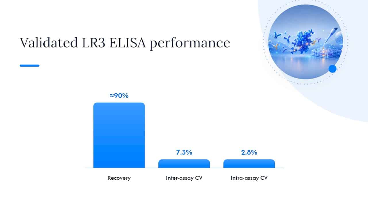
The most reliable option identified in the literature is an LR3-specific ELISA built around an antibody targeting the Arg3 epitope, which native IGF-1 doesn’t carry. This design lets the assay discriminate LR3 from endogenous IGF-1 and IGF-2 directly, rather than relying on extraction steps to strip out interfering binding proteins. The validated version of this assay, described in the original ELISA development paper, detected as little as 50 pg of LR3 in a 100 μL sample, with an intra-assay coefficient of variation of 2.8% and an inter-assay CV of 7.3%, and recovery around 90%. Critically, the assay’s design makes it robust to IGFBP interference, which is the single biggest source of error in older radioimmunoassay (RIA) approaches.
RIA-based methods can still work, but only with extra sample preparation. Because IGFBPs bind IGF-1 and IGF-1 analogues with high affinity, an RIA run on raw plasma will underestimate free ligand and produce inconsistent results depending on each sample’s IGFBP load. Acid extraction or size-exclusion chromatography prior to RIA strips out the bound fraction and gives a more accurate total measurement, but adds processing time and a real risk of variable recovery between batches.
For labs setting up LR3 quantification, a few practical steps head off the most common failure modes:
- Run a dilution series on every new sample matrix (plasma, serum, cell culture media) before trusting single-point readings, since matrix effects vary by IGFBP content.
- Include spike-recovery checks with known LR3 concentrations added to your actual sample matrix, not just assay buffer, to confirm the ~90% recovery benchmark holds in your hands.
- Report intra- and inter-assay CVs alongside your data, using the published 2.8% and 7.3% figures as your validation target.
- State explicitly whether your protocol used the LR3-discriminating ELISA or a generic IGF assay with extraction, since these are not interchangeable methods.
Pro Tip: Always report the IGFBP status of your sample matrix alongside your LR3 concentration data. A concentration measured in IGFBP-depleted media means something functionally different from the same number in whole plasma, and burying that detail in methods makes your dose-response curve nearly impossible for another lab to replicate.
What Systemic Effects Should Researchers Monitor for Safety?
The systemic endocrine response to LR3 exposure is not a minor side effect to footnote. It’s often the dominant signal in the dataset, and treating it as background noise is how researchers end up misreading their own results.
In the porcine infusion study, the systemic picture was unambiguous: a 4-day exposure at 180 μg/kg/day mean plasma growth hormone was markedly reduced, the GH peak area under the curve was greatly decreased, and plasma IGFBP-3, endogenous IGF-1, and insulin levels also declined. That’s four interconnected axes moving together in response to a single intervention, which tells you the somatotropic feedback loop reacts to LR3 exposure as a strong, coordinated signal rather than a localized nudge.
The magnitude of that GH suppression, a 23% drop in mean concentration and a 60% drop in peak AUC from just four days of infusion, is large enough that it should be a primary endpoint in any LR3 protocol, not an incidental finding buried in a supplementary table.
The fetal-sheep work adds a second dimension to the safety picture: metabolic substrate effects independent of the classic GH axis. That study found no change in glucose-stimulated insulin secretion or glucose and oxygen levels, but circulating amino acids dropped significantly. In a growth-restricted fetus already operating under substrate constraint, pulling amino acids down further is a meaningful signal, not a side note, particularly given that the intervention produced no compensating growth benefit to justify the metabolic cost.
Taken together, these two studies point toward a consistent monitoring panel for any LR3 protocol, whether the goal is mechanistic characterization or a translational feasibility test:
- Growth hormone, sampled with attention to timing relative to infusion, since both mean concentration and pulsatile peak structure shifted in the pig data.
- Insulin and glucose, to catch metabolic shifts even when growth outcomes look neutral.
- IGFBP-3, as a marker of endogenous IGF axis suppression rather than just an interference variable in your assay.
- Circulating amino acids, especially in any nutrient-limited or growth-restriction model, given the significant drop observed in the fetal-sheep data.
There’s also an animal welfare and translational ethics dimension that deserves direct acknowledgment. The fetal-sheep model showing decreased amino acids with no offsetting growth benefit is a harm signal without a compensating gain, which is exactly the kind of result that should slow down, not accelerate, any push toward applying LR3 in growth-restriction contexts before the mechanism is better understood. Boosting receptor-available signaling doesn’t help much when the underlying constraint is substrate or oxygen availability rather than receptor occupancy. Designing safety monitoring around this GH/insulin/IGFBP-3 axis, rather than isolated tissue-level readouts, is the more defensible approach given what the pig and fetal-sheep data actually show.
Where Does LR3 Show Real Therapeutic Promise, and Where Doesn’t It?
The honest answer sits between two extremes: LR3 has a defensible mechanistic case for muscle-related research applications, and a documented failure case in nutrient-limited growth models. Both matter, and conflating them is where a lot of secondhand summaries go wrong.
On the supportive side, IGF-1 signaling has a well-established role in muscle repair and hypertrophy pathways, working through satellite cell activation and protein synthesis machinery. The review on optimizing IGF-1 for skeletal muscle therapeutics frames the therapeutic rationale clearly while also flagging that the field hasn’t solved the practical problems: which IGF-1 form to use, how to manage extracellular matrix storage and release, and how to modulate receptor availability without triggering the kind of systemic feedback seen in the pig study. LR3’s low IGFBP affinity is theoretically attractive here because it could mean more sustained local receptor engagement in tissue, but that same property is exactly what drove the systemic GH suppression documented in vivo.
The null result side is just as important to weigh. The fetal-sheep growth-restriction study is the clearest available evidence that increasing receptor-available IGF signaling doesn’t automatically translate into a growth benefit when the underlying limitation is nutrient or oxygen supply rather than receptor occupancy. That’s a mechanistic ceiling, not a dosing problem; more LR3 or a different infusion schedule likely wouldn’t fix it, because the bottleneck sits upstream of the receptor entirely.
Key evidence gaps that limit any confident translational claim right now include the following:
- No published multi-week chronic exposure data exists for LR3 in a mammalian model, so long-term safety and adaptation effects are unknown.
- Optimal dosing windows haven’t been established outside the two infusion-based studies reviewed here, and bolus versus continuous administration hasn’t been directly compared.
- Systemic endocrine interactions (GH axis suppression specifically) haven’t been characterized across a dose-response curve, only at single doses in each study.
- Muscle-tissue-specific pharmacokinetics for LR3, as distinct from plasma pharmacokinetics, remain largely uncharacterized in the published literature.
| Application area | Evidence strength | Main limitation |
|---|---|---|
| Muscle repair/hypertrophy mechanism | Mechanistically plausible, supported by review-level synthesis | No LR3-specific chronic dosing trial published |
| Fetal growth restriction rescue | Tested directly, negative result | Substrate-limited model, no growth benefit, reduced amino acids |
| Systemic endocrine safety profile | Documented in two species, consistent suppression signal | Short exposure windows only; chronic data absent |
The responsible experimental path forward is to design studies that isolate the variable you actually care about. If the goal is tissue-level signaling, pair LR3 exposure with local delivery or ex vivo tissue models to minimize systemic GH-axis confounds. If the goal is a translational feasibility question, build in the same endocrine monitoring panel used in the pig and fetal-sheep studies from day one, rather than adding it retroactively once an unexpected result shows up.
Why Reagent Quality Determines Whether Your LR3 Data Is Reproducible
Every conclusion in this article rests on the assumption that the LR3 used in each study was what the label said it was. That assumption breaks down fast with poorly characterized research peptides, which is why Data Peptides treats Certificate of Analysis (COA) documentation as a non-negotiable part of sourcing LR3 for laboratory work.
A COA worth trusting reports several specific data points, not just a purity headline:
- Purity percentage, ideally 99% or higher, verified by HPLC or mass spectrometry rather than a vendor’s internal estimate alone.
- Identity confirmation, matching the peptide’s mass and sequence to the expected LR3 structure.
- Endotoxin levels, particularly important for any in vivo or cell culture application where contamination could confound endocrine or inflammatory readouts.
- Storage recommendations, since peptide stability depends heavily on temperature and reconstitution handling.
Third-party testing matters here because it removes the vendor as the sole judge of its own product. An independent lab verifying purity and identity catches batch-to-batch variance that could otherwise show up in your data as unexplained noise between replicate experiments, particularly relevant given how sensitive LR3’s apparent potency already is to context. Data Peptides publishes COA documentation on its IGF-1 LR3 product page for exactly this reason, and pairs it with laboratory handling standards covering reconstitution and storage protocols that affect peptide integrity between receipt and use.
One more point worth stating plainly: LR3 sold for research is labeled research-use-only, and that labeling exists for a reason. Institutional compliance requirements around handling and use apply regardless of how the peptide is sourced, and any lab bringing LR3 into a protocol should confirm its use falls within the appropriate institutional and regulatory framework before ordering.
What This Research Actually Tells Us About LR3
The clearest lesson from the LR3 literature is that receptor-level signaling and whole-organism physiology don’t move in lockstep, and treating LR3 as a simple “more potent IGF-1” is a category error the Francis et al. data disproved decades ago. Potency isn’t a fixed number here; it’s a function of the IGFBP environment you’re testing in, and ignoring that turns a mechanistic tool into a source of contradictory data.
The two anchor animal studies, spanning postnatal pigs and growth-restricted fetal sheep, tell a consistent story about systemic feedback overriding local receptor availability. Any research program built on LR3 should treat GH-axis suppression as a primary variable to measure, not a confound to explain away after the fact. Study designs that isolate tissue-level effects from systemic ones, through local delivery, ex vivo models, or tightly controlled endocrine monitoring, will produce far more interpretable data than a systemic dose escalation alone.
For labs building out that kind of protocol, sourcing matters as much as design. Data Peptides maintains COA-backed, third-party-tested LR3 specifically so that reagent variability doesn’t become another uncontrolled variable in an already context-sensitive system.
— Data Peptides
Sourcing Research-Grade LR3 for Reproducible Work
Every point in this article assumes the LR3 in your protocol is exactly what the label claims, and that’s the gap Data Peptides exists to close. Unlike sourcing routes where purity documentation is vague or missing entirely, Data Peptides pairs every LR3 batch with third-party testing and a Certificate of Analysis covering purity, identity, and endotoxin data, so batch variance doesn’t quietly distort your dose-response curves.
That matters most for researchers running the kind of context-sensitive protocols this article describes, where an unverified peptide source can introduce noise indistinguishable from a real biological signal. Data Peptides ships fast and discreetly within the US, backs every order with 24/7 support, and labels every product clearly for research-use-only applications.
If you’re setting up an LR3 protocol and need a reagent source you can actually document in your methods section, review the current IGF-1 LR3 listing and check current COA data before your next order.
This article is general information, not a substitute for advice from a qualified doctor. Consult a qualified healthcare professional about your own circumstances before acting on anything here.
Sources
- Long R3 insulin-like growth factor-I reduces growth, plasma growth hormone, IGF binding protein-3 and endogenous IGF-I concentrations in pigs
- IGF-1 LR3 does not promote growth in late-gestation growth-restricted fetal sheep | American Journal of Physiology-Endocrinology and Metabolism
- Novel recombinant fusion protein analogues of insulin-like growth factor (IGF)-I indicate the relative importance of IGF-binding protein and receptor binding for enhanced biological potency
- Optimizing IGF-I for skeletal muscle therapeutics
FAQ
Does IGF-1 LR3 Actually Work?
It reliably activates the IGF-1 receptor and behaves differently depending on IGFBP context, but the two major animal studies to date show it does not straightforwardly improve growth outcomes and can suppress the body’s own GH and IGF-1 production.
Is IGF-1 LR3 Stronger Than HGH?
They aren’t directly comparable. LR3 acts on the IGF-1 receptor while HGH works upstream to stimulate endogenous IGF-1 production, and the pig study found that co-administering GH alongside LR3 produced no additional growth effect, suggesting the two don’t simply stack.
Does IGF-1 LR3 Grow Muscle?
Mechanistic reviews support a plausible role for IGF-1 signaling in muscle repair and satellite cell activation, but no published LR3-specific study has demonstrated a clear muscle growth benefit in a controlled, chronic-dosing animal model.
How Should IGF-1 LR3 Dosage Be Reported in Research?
Published studies used continuous infusion at 180 μg/kg/day for four days in pigs and about 1.17 μg·kg⁻¹·h⁻¹ for one week in fetal sheep, and any new protocol should report exact units, infusion rate, and IGFBP status of the sample matrix for comparability.