

Physician-directed PTH analogs like teriparatide and BMP-based bone grafting have the strongest clinical evidence behind them for supporting fracture repair. Below that top tier sits a group of engineered peptides, Ab46-D-Glu20, PEPITEM, TP508, and PepGen P-15, with genuinely encouraging preclinical data but no large human fracture trials yet. Everything else on the “peptides for bone healing” list you’ve probably seen (OGP, RGD, GFOGER, thymosin beta-4, BPC-157) sits further back, backed mostly by cell culture and small-animal work.
That distribution matters for how you read the literature. A 2016 systematic review covering dozens of in vitro and animal studies found that many peptides upregulate markers of bone healing, but clinical trial evidence remains limited for most of them. The gap between “works in a mouse” and “works in a person with a tibial fracture” is wide, and it’s usually a delivery problem, not a biology problem.
What the evidence actually supports right now:
- Teriparatide and BMP-2/BMP-7 have documented clinical use, with real limitations around dosing, cost, and ectopic bone risk.
- Ab46-D-Glu20, a fracture-targeted PTHrP-like construct, cut fracture repair time by more than half and roughly doubled mechanical strength in preclinical mouse and dog models.
- PEPITEM shows dual osteoblast-stimulating and osteoclast-inhibiting activity in preclinical disease models, a mechanism profile researchers rarely see in a single molecule.
- Most other candidates, including BPC-157 and thymosin beta-4, remain exploratory, with signal in rodents but little to no controlled human fracture data.
If you’re designing a study or sourcing material for lab work, the practical takeaway is this: match your expectations to the evidence tier, prioritize delivery and retention in your protocol, and confirm sourcing with a verified Certificate of Analysis before you start.
Key Takeaways
The strongest evidence for peptides in bone healing sits with physician-directed PTH analogs and surgical BMP applications, while engineered candidates like Ab46-D-Glu20 and PEPITEM lead the preclinical field.
| Point | Details |
|---|---|
| Clinical tier is narrow | Only teriparatide and BMP-2/BMP-7 have meaningful human fracture-relevant data; both carry real dosing and safety limitations. |
| Ab46-D-Glu20 leads preclinical targeting | Fracture-targeted delivery cut repair time by over half and nearly doubled mechanical strength in animal models. |
| Dual-action peptides stand out | PEPITEM’s ability to stimulate osteoblasts and inhibit osteoclasts simultaneously is mechanistically rare among bone-healing peptides. |
| Delivery is the real bottleneck | Rapid plasma clearance, not weak biology, explains why many promising peptides underperform in vivo without targeting or carrier systems. |
| Verify sourcing before research | Confirm Certificates of Analysis and third-party purity testing, resources like Data Peptides’s COA page support that verification step. |
Table of Contents
- Which Peptides Have Evidence for Bone Healing?
- How Peptides Actually Trigger Bone Repair
- Why Delivery Determines Whether a Peptide Works
- What Human Trials Show, and Where the Data Runs Out
- Designing Reproducible Peptide Research for Bone Repair
- Sourcing Research Peptides for Bone Healing Studies
- Side Effects and Contraindications Researchers Should Track
- Integrating Peptide Research with Surgical and Rehabilitation Protocols
- Peptides for Bone Healing: Evidence and Mechanisms for Researchers
- Research-Grade Peptides for Your Bone Healing Studies
- Sources
- FAQ
Which Peptides Have Evidence for Bone Healing?
Sorting bone healing peptides by evidence tier keeps you from treating a cell-culture finding and a human trial result as equivalent proof. Here’s how the field actually breaks down, from strongest human data to weakest.
Clinical and physician-directed: teriparatide and BMP-based approaches
Teriparatide, a recombinant fragment of parathyroid hormone, has the most mature human data of anything on this list. It’s approved for osteoporosis and used off-label by some physicians to support fracture healing, particularly in patients with delayed union. It works by stimulating osteoblast activity when given intermittently, a dosing nuance that matters: continuous PTH exposure favors bone resorption, while pulsed dosing favors formation.
BMP-2 and BMP-7 occupy a different niche. These aren’t systemic drugs; they’re applied locally, usually on a collagen sponge, during spinal fusion or open fracture surgery. The BMC Medicine review on peptides in bone regeneration notes that BMP-derived peptides are among the most studied bone-healing agents in preclinical and clinical settings, yet translating BMP-mimetic peptides into consistently reliable therapies has proven harder than the early data suggested. Dosing windows are narrow, and ectopic bone formation, essentially bone growing where you don’t want it, remains a documented complication.
Evidence level: Human clinical use, established but with real limitations.
Implication for research: Useful as a benchmark for effect size when evaluating newer candidates, but not a template to copy without addressing the same dosing and safety issues.
Strong preclinical candidates with translational momentum
This tier is where the most interesting bone repair peptide research is happening right now.
Ab46-D-Glu20 is a fracture-targeted construct that fuses a PTHrP-like anabolic payload to a D-Glu20 sequence, which binds selectively to exposed hydroxyapatite at fracture surfaces. In mouse models, the targeted version achieved more than 12 times the cumulative residence time at the fracture site compared to the untargeted peptide alone, cut repair time by over half, and nearly doubled final mechanical strength, according to the engineered dual-function peptide study. That targeting strategy also worked in dogs, which matters because canine bone remodeling rates are closer to human physiology than rodent models.

PEPITEM is an endogenous osteopeptide that does two things most single-target drugs can’t: it stimulates osteoblast maturation and new bone formation while triggering osteoprotegerin (OPG) release, which suppresses osteoclast activity. The PEPITEM study documented increased alkaline phosphatase activity and enhanced mineralization alongside OPG-mediated inhibition of bone resorption in preclinical models. Acting on both sides of the remodeling equation, rather than just formation or just resorption, is closer to how romosozumab works clinically, except PEPITEM gets there through an endogenous peptide pathway instead of a monoclonal antibody.
TP508, also known as Chrysalin, is a synthetic fragment of the thrombin receptor-binding domain. Preclinical work links it to improved fracture callus formation and enhanced angiogenesis at the repair site, two processes that typically move in tandem during bone healing. It’s been studied in diabetic fracture models specifically, where impaired vascular response is a known barrier to healing.
PepGen P-15, a synthetic 15-amino-acid peptide derived from type I collagen, has clinical traction, but in a narrower lane than teriparatide or BMP products. It shows real utility in dental and maxillofacial bone graft applications, where defect size and mechanical load are modest. The systematic review on peptides in bone regeneration notes that extrapolating this data to large long-bone non-unions requires caution, since the mechanical and scale demands of a femoral fracture are nothing like a dental socket.
Evidence level: Robust animal data, in some cases across two species; no large-scale human fracture trials yet.
Implication for research: These are the candidates worth prioritizing if you’re planning translational or mechanistic follow-up studies, precisely because the preclinical foundation is unusually solid.
Exploratory candidates: promising signals, real evidence gaps
A cluster of peptides shows up constantly in bone healing discussions despite having thinner human data than either tier above.
- OGP (osteogenic growth peptide): A naturally occurring serum peptide shown in vitro to stimulate osteoblast proliferation; animal fracture data exists but is limited in scope.
- RGD-containing peptides: Short sequences (arginine-glycine-aspartate) that mimic cell-adhesion motifs found in fibronectin and other extracellular matrix proteins; used mainly as surface coatings on implants rather than as standalone therapeutics.
- GFOGER: A collagen-derived peptide sequence that binds integrin receptors on osteoblast precursors, studied mostly for its role in scaffold and biomaterial design rather than systemic or local injection therapy.
- Thymosin beta-4: Has direct mouse fracture-healing data showing improved outcomes, according to a review of fracture repair research, but human fracture trials are absent. Data Peptides covers the underlying biology on its thymosin beta-4 resource page.
- BPC-157: Popular in research circles for tissue repair broadly, with multiple animal-model signals for bone and soft tissue, but limited human evidence and FDA safety flags around compounded, unregulated versions. Data Peptides’s BPC-157 and TB-500 resource page breaks down the mechanism data researchers cite most often.
- Collagen-derived peptides: Widely used in supplement and scaffold contexts; evidence for direct fracture-repair acceleration in humans is sparse compared to their role as structural scaffolding material.
Evidence level: In vitro and small-animal studies predominate; human fracture-repair data is either absent or anecdotal.
Implication for research: Useful for mechanistic study or as scaffold components, but claims about “accelerating bone healing” in humans outpace what the current data can support.
How Peptides Actually Trigger Bone Repair
Bone repair peptides don’t all work the same way, and knowing which mechanism a candidate targets tells you what to measure when you evaluate it. Fracture healing runs through four overlapping processes: inflammation, soft callus formation, hard callus mineralization, and remodeling. Different peptides intervene at different points.
Osteoblast activation is the most common target. Peptides that upregulate alkaline phosphatase (ALP) activity, RUNX2 expression, and downstream mineralization are pushing precursor cells toward mature, bone-forming osteoblasts. BMP-derived motifs work this way directly, engaging the BMP receptor pathway. OGP appears to work through a related but distinct route, stimulating osteoblast proliferation in culture, though the exact receptor mechanism is still debated.
Osteoclast regulation runs through the OPG/RANKL axis, the signaling system that governs how aggressively bone gets broken down during remodeling. RANKL promotes osteoclast formation; OPG blocks it by acting as a decoy receptor. PEPITEM is the clearest example of a peptide working both sides at once: it stimulates osteoblast maturation while independently triggering OPG release to rein in osteoclast activity. Most peptides only touch one side of that balance, which is what makes PEPITEM’s dual mechanism worth watching closely.
Angiogenesis and vascular coupling matter more than most non-specialists appreciate. Bone doesn’t heal without a blood supply feeding the repair site, and the coupling between new vessel formation and new bone formation is tight enough that researchers now treat them as a single process rather than two. TP508’s effect on callus formation appears to run partly through enhanced angiogenesis, which may explain why it’s been studied specifically in diabetic models, where vascular response is often impaired.
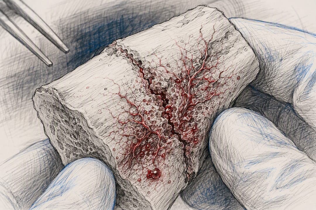
Matrix-mimetic peptides work through an entirely different logic. RGD sequences mimic the cell-adhesion motifs found in fibronectin and other extracellular matrix proteins, giving osteoblast precursors something to grip onto. GFOGER does the same for collagen-derived integrin binding. Neither peptide “signals” cells to differentiate the way BMP or OGP do; instead, they create a physical and biochemical surface that supports adhesion and, downstream, mineral nucleation. That’s why you’ll find RGD and GFOGER coating implants and scaffolds far more often than you’ll find them injected as standalone therapeutics.
Pro Tip: If you’re screening a new peptide for osteogenic potential, run ALP activity and mineralization assays (Alizarin Red staining is standard) alongside a RUNX2 expression check. A peptide that boosts one but not the others is telling you something about which stage of osteoblast differentiation it’s actually influencing, not just whether it “works.”
Why Delivery Determines Whether a Peptide Works
Most peptides that look promising in a test tube fail somewhere between the bench and the bedside, and it’s almost never because the peptide lacks biological activity. It’s because peptides are fragile. They get cleared from plasma within minutes to hours, degraded by circulating proteases before they ever reach meaningful concentration at a fracture site. A molecule that stimulates osteoblasts beautifully in culture is useless if it’s gone from the bloodstream before it finds the injury.
The Ab46-D-Glu20 construct solves this with a targeting strategy rather than a formulation trick. By fusing the anabolic peptide to a D-Glu20 sequence that binds exposed hydroxyapatite, researchers achieved dramatically longer residence time specifically at the fracture surface, more than 12 times the cumulative exposure compared to the untargeted version, while systemic exposure elsewhere in the body stayed comparable. That’s a meaningful distinction: the peptide can be dosed systemically, through an IV or subcutaneous injection, yet its anabolic effect concentrates almost entirely where the bone is actually broken. Fewer off-target effects, better local dosing, in a single design choice.
Where targeting ligands aren’t built into the peptide itself, researchers lean on carrier systems instead. A review of nanoengineered therapeutic peptides lays out the main options:
- Hydrogels provide a soft, injectable matrix that holds peptides at a local site and releases them gradually, useful when you need sustained exposure over days rather than a single burst.
- Peptide amphiphiles self-assemble into nanofibers that mimic the structure of native extracellular matrix, giving cells a scaffold to adhere to while presenting bioactive peptide sequences on the surface.
- Mesoporous carriers (silica or calcium-phosphate based) load peptides into a porous structure that controls release kinetics and can be tuned for faster or slower diffusion depending on the healing stage being targeted.
- Self-assembling nanofiber systems organize at the injury site to present peptide signals in a spatially structured way, closer to how growth factors are actually displayed during natural bone repair.
| Delivery approach | Best suited for | Key limitation |
|---|---|---|
| Fracture-targeting ligands (D-Glu20) | Systemic dosing with local concentration | Requires peptide engineering, not a drop-in fix |
| Hydrogels | Sustained local release over days | Limited to accessible or surgically exposed sites |
| Peptide amphiphiles | Scaffold-integrated signaling | More complex manufacturing |
| Mesoporous carriers | Tunable, staged release | Loading capacity varies by peptide size |
If you’re designing an experiment, the most informative comparison isn’t peptide A versus peptide B in isolation. It’s local application versus systemic targeted dosing of the same peptide, measured against both residence time at the fracture site and functional outcomes like mechanical strength and radiographic healing. A peptide that shows strong in vitro activity but poor retention in vivo is a delivery problem waiting for an engineering solution, not a failed candidate.
What Human Trials Show, and Where the Data Runs Out
Teriparatide has the most human outcome data of any peptide discussed here, largely from osteoporosis trials that included fracture-healing endpoints as secondary measures. Benefits show up most consistently in delayed union cases and in patients with reduced bone density to begin with; the effect size in otherwise healthy fracture patients is less consistently demonstrated. BMP-2 and BMP-7 have a similar profile: strong evidence in spinal fusion and open tibial fracture contexts, weaker or absent evidence outside those specific surgical indications.
Beyond that, the human data thins out fast. There are no large controlled human fracture-healing trials for BPC-157 or thymosin beta-4. Everything cited for these two peptides, including direct mouse fracture-healing improvements for thymosin beta-4, comes from animal models. That’s not a knock on the biology; mouse and rat fracture models are standard, validated tools in orthopedic research. It just means anyone citing these peptides as proven human fracture-healing agents is overstating the evidence.
Safety and regulatory flags worth knowing:
- BMP products carry a documented risk of ectopic bone formation and, in some surgical contexts, increased local inflammation.
- Some peptide classes raise immunogenicity concerns, particularly with repeated systemic dosing over time.
- BPC-157 specifically carries FDA cautions related to compounded, unregulated formulations, a distinct issue from its preclinical biology.
- Off-label or compounded peptide use for fracture healing sits in a regulatory gray zone in most jurisdictions and should never be treated as equivalent to an approved therapeutic.
Before any peptide moves toward clinical translation, the standard path runs through GLP (Good Laboratory Practice) toxicology studies, dose-escalation trials starting at the lowest anticipated biologically active dose, and institutional review board approval for any human-adjacent work. Skipping that sequence isn’t a shortcut; it’s how promising preclinical candidates turn into safety liabilities.
Designing Reproducible Peptide Research for Bone Repair
Getting usable data out of a bone-healing peptide study starts before the first animal or cell plate, at the sourcing stage. A peptide with an unconfirmed sequence or unlisted purity level will confound your results no matter how well-designed the rest of the protocol is.
- Verify sourcing before you start. Request third-party purity testing, a full Certificate of Analysis, mass spectrometry sequence confirmation, and endotoxin testing for anything intended for cell culture or in vivo work. Maintain chain-of-custody documentation so results are traceable back to a specific batch.
- Match dosing context to your research question. Translating an animal exposure level to a human-relevant dose isn’t a simple weight-based conversion; metabolic rate and clearance kinetics differ meaningfully between species. Choose local versus systemic administration based on the mechanism you’re testing, a targeting-ligand study needs systemic dosing to demonstrate its advantage, while a scaffold-integration study calls for local delivery from the start.
- Select outcome measures that match the fracture model’s timeline. Standard measures include radiographic union scoring, histological analysis of callus composition, biomechanical testing (torsion or bending strength), and angiogenesis markers like CD31 staining. Early time points capture inflammation and soft callus formation; later time points are needed to assess mineralization and mechanical strength.
- Build in the controls that make results defensible. Carrier-only control groups rule out delivery-vehicle effects. Blinded scoring for radiographic and histological outcomes removes observer bias. Power your study for the endpoint that matters most, mechanical strength, not just the easiest one to measure, radiographic appearance.
Pro Tip: Report raw data alongside your Certificate of Analysis when publishing or sharing results. A surprising number of conflicting findings in the peptide literature trace back to unverified peptide purity rather than genuine biological variability.
Data Peptides’s general peptide research resource covers additional context on lab-use applications if you’re scoping a new project.
Sourcing Research Peptides for Bone Healing Studies
Reproducible results depend on knowing exactly what’s in the vial. Data Peptides supplies lab-grade peptides with documentation built for that standard, not an afterthought bolted onto a product listing.
- Every peptide ships with a third-party Certificate of Analysis confirming purity, typically at 99% or above, and sequence accuracy.
- COAs are available for review before purchase through the company’s Certificate of Analysis page, so you can verify a batch before it’s part of your protocol.
- Product-specific resource pages, including coverage of thymosin beta-4 and BPC-157, summarize the mechanism data referenced throughout this article.
- The broader peptides used for research overview outlines testing policies and lab-use guidance across the product catalog.
Everything Data Peptides sells is intended for laboratory research use only. None of it is manufactured, labeled, or distributed as a medical treatment, and nothing here should be read as a substitute for physician-directed care. Researchers working with any of the peptides discussed in this article should confirm institutional approval and appropriate handling protocols before beginning work.
Side Effects and Contraindications Researchers Should Track
Adverse findings in the bone-healing peptide literature cluster around a few recurring issues. BMP-based products are associated with ectopic bone formation, swelling, and localized inflammatory response, effects that show up specifically at the surgical application site rather than systemically. Peptides administered repeatedly over extended periods raise a separate concern: immunogenicity, where the body develops antibody responses that can blunt the peptide’s effect over time or trigger localized reactions.
Compounded and unregulated peptide products carry their own risk profile, distinct from the underlying biology. BPC-157’s regulatory flags stem specifically from concerns about compounded formulations lacking consistent quality control, not from a documented human safety signal in controlled trials, because those controlled trials largely don’t exist yet.
Contraindications in animal models offer some early guidance, even without human trial data to confirm them. Peptides that stimulate aggressive osteoblast activity warrant caution in contexts involving active malignancy, given the theoretical concern about stimulating abnormal cell proliferation in bone. Cardiovascular signals have also been flagged for select peptide classes in preclinical safety screening, an area that needs closer scrutiny before any human translation work proceeds.
For any research protocol involving live animal models or cell lines derived from patients, safety monitoring should track both the intended osteogenic endpoint and off-target markers, liver and kidney function panels at minimum, to catch systemic effects that a narrow focus on bone outcomes alone would miss.
Integrating Peptide Research with Surgical and Rehabilitation Protocols
Peptides don’t replace surgical fixation, physical therapy, or standard fracture management; in every clinical context where they’re used, they’re additive. Teriparatide, for example, is typically layered onto standard fracture care rather than substituted for it, and BMP products are applied directly during surgical fixation, not as a standalone intervention.
That integration principle matters for research design too. A peptide that shows strong osteogenic activity in isolation may perform differently when tested alongside mechanical loading, since bone responds to mechanical stress through its own signaling pathways (the piezoelectric effect and Wolff’s Law dynamics), which can either synergize with or compete against a peptide’s biochemical signal depending on timing.
Practical integration points to build into a translational research plan:
- Surgical timing: If a peptide is meant for local application, it needs a delivery vehicle (a bone graft substitute, collagen sponge, or hydrogel) that survives the surgical environment and integrates with standard fixation hardware.
- Rehabilitation timing: Early mechanical loading protocols in physical therapy influence callus remodeling, and studies testing peptide efficacy without controlling for rehabilitation timing risk confounding a peptide effect with a loading effect.
- Sequencing with pharmacologic care: Patients already on bisphosphonates or other bone-active medications introduce a confound that needs explicit control arms, since these drugs affect the same OPG/RANKL and remodeling pathways many peptides target.
Any protocol combining a research peptide with a surgical or rehabilitation intervention needs its own institutional review, since safety and efficacy data rarely exist for the combination even when they exist for the peptide alone.
Peptides for Bone Healing: Evidence and Mechanisms for Researchers
The biggest misreading in this field is treating “peptide” as one category with one evidence level. It isn’t. Teriparatide belongs in a fundamentally different conversation than BPC-157, and lumping them together, as most consumer-facing content does, does researchers a disservice.
The conventional framing also undersells delivery. Most write-ups focus entirely on which peptide does what to which cell type, and treat pharmacokinetics as an afterthought. But the Ab46-D-Glu20 data makes a strong case that targeting and retention engineering deserve equal billing with the peptide’s core biology. A mediocre peptide with excellent local retention will likely outperform a superior peptide that clears the bloodstream in twenty minutes.
If you take one thing from this article, prioritize evidence tier and delivery mechanism over novelty. The peptides worth your attention right now are the ones where researchers have already solved, or are actively solving, the retention problem.
Research-Grade Peptides for Your Bone Healing Studies
Reliable bone-healing peptide research depends on knowing exactly what’s in every vial, and that’s the gap Data Peptides was built to close. Fast, discreet US shipping and a 24/7 support team mean you’re not stuck waiting on sourcing questions mid-protocol. If you’re planning a study involving any of the candidates covered here, start by reviewing the research-grade peptide catalog and confirm purity documentation before your next order. All products are strictly for laboratory research use.
This article is general information, not a substitute for advice from a qualified doctor. Consult a qualified healthcare professional about your own circumstances before acting on anything here.
Sources
- The role of peptides in bone healing and regeneration: a systematic review
- PEPITEM (endogenous osteopeptide) study
FAQ
What peptides are good for bone repair?
Teriparatide and BMP-2/BMP-7 have the strongest clinical evidence, while Ab46-D-Glu20, PEPITEM, TP508, and PepGen P-15 show the most promising preclinical data for bone regeneration.
Is BPC-157 good for broken bones?
BPC-157 has multiple animal-model signals for tissue and bone repair, but human fracture-healing trial data is absent, and compounded formulations carry FDA safety cautions.
What’s the downside of taking peptides?
Documented concerns include ectopic bone formation with BMP products, immunogenicity with repeated dosing, and quality-control risks tied to unregulated or compounded peptide sources.
How can I speed up healing of a broken bone?
Standard fracture care, proper fixation, adequate nutrition, and physical therapy, remains the foundation; peptide-based approaches like teriparatide are used as additive therapy under physician direction, not as replacements for standard care.
Are peptides for bone healing approved for human use?
Only a small subset, teriparatide and BMP-2/BMP-7 among them, have established clinical use; most peptides discussed in current research, including thymosin beta-4 and BPC-157, remain preclinical and are not approved fracture treatments.
This article is intended for informational and research purposes only and does not constitute medical advice. Consult a qualified physician before making any decisions about fracture treatment or peptide use in a clinical context.